Sunuyu indir
Sunum yükleniyor. Lütfen bekleyiniz

1
Moleküler Testlerde Preanalitik Faz
Prof. Dr. Abdulkerim BEDİR Ondokuz Mayıs Üniversitesi Samsun 2014

2
Lab Döngüsü

3
Biyospesmen and süreç Biyospesmen Bilgilerinin Derleme ve Analizi
Hasta Edinim/ Acquisition İşlem/ Proses Saklama Dağıtım Bilimsel Analiz Tıbbi/ Cerrahi Prosedürler Artanı Stoklama Biyospesmen Bilgilerinin Derleme ve Analizi Bilimsel Bilgilerin Derleme ve Analizi Klinik/Klinik Araştırma Çıktıları

4
Biyospesmenlere bağlıdır
Moleküler Amaç Genomics Proteomics Metabolomics 1-Teşhis etmek 2-Tedavi belirlemek 3-Risk analizi Yüksek kalitede Biyospesmenlere bağlıdır

5
Moleküler İdeal Bugün Yarın
Three colon cancer patients: Same disease? Same therapy? Each patient differs with respect to the molecular basis of his/her cancer

6
Moleküler Evren

7
Moleküler Doğma

8
Moleküler Akış

9
Preanalitik faktörler I: Sıvı Biyospesmen
Antikoagülanlar Sitrat-DNA, RNA EDTA-DNA Heparin-Sitolojik testler Stabilizör/İnhibitörler Protein: Proteaz inhibitörleri RNA: Beta-merkaptoetanol (stabilizör) RNaz inhibitörleri DNA: Stabildir-Örn. Guthrie testi Saklama sıcaklığı PMSF, Leupeptin, pepstatin, aprotinin 2-Mercaptoethanol is used in some RNA isolation procedures to eliminate ribonuclease released during cell lysis. Numerous disulfide bonds make ribonucleases very stable enzymes, so 2-mercaptoethanol is used to reduce these disulfide bonds and irreversibly denature the proteins. This prevents them from digesting the RNA during its extraction procedure
 RNaz inhibitörleri. DNA: Stabildir-Örn. Guthrie testi. Saklama sıcaklığı. PMSF, Leupeptin, pepstatin, aprotinin. 2-Mercaptoethanol is used in some RNA isolation procedures to eliminate ribonuclease released during cell lysis. Numerous disulfide bonds make ribonucleases very stable enzymes, so 2-mercaptoethanol is used to reduce these disulfide bonds and irreversibly denature the proteins. This prevents them from digesting the RNA during its extraction procedure.")
10
Endojen bozucu etkenler
Bekleme süresi Hücre sayımı 24 saatte düşer Sterilite Bakteriyal kontaminasyon Fungal kontaminasyon Endojen bozucu etkenler Enzimler: Proteazlar, DNazlar, RNazlar Hücre ölümü Numune tüpleri/kapları DNaz-free RNaz-free Steril

11
Antikoagulanlar Aditif Renk Testler Yok Kırmızı
Biyokimya, Seroloji, immünoloji, serum Sodium heparin (freeze dried) Yeşil Immünoloji, viroloji testleri Sodium heparin Kahverengi Sitogenetik testler, moleküler testler Tripotassium EDTA (7.5-15% solution) Mor Virology, molecular biology Acid citrate dextrose (ACD) solution Sarı Moleküler biyoloji
 Yeşil. Immünoloji, viroloji testleri. Sodium heparin. Kahverengi. Sitogenetik testler, moleküler testler. Tripotassium EDTA (7.5-15% solution) Mor. Virology, molecular biology. Acid citrate dextrose (ACD) solution. Sarı. Moleküler biyoloji.")
12
Preanalitik faktörler II: Solid Biyospesmen
Kanser hücrelerinde genomik değişiklikler düşük frekanstadır Kantite sorunu Biyopsi materyali Kalite sorunu Biyopsi materyalleri formalinle fiksedir. DNA izolasyonu özel protokole tabidir Pürite sorunu (Tümör heterogenitesi) Normal hücreler ile karışıktır Kanserin kendisinde heterogenite (farklı klonlar) RNAlater RNA Stabilization Reagent immediately stabilizes RNA in tissue samples to preserve the gene expression profile, and is supplied in 50 ml or 250 ml bottles. For stabilization of DNA, RNA, and protein in tissue samples, Allprotect Tissue Reagent can be used.
 Normal hücreler ile karışıktır. Kanserin kendisinde heterogenite (farklı klonlar) RNAlater RNA Stabilization Reagent immediately stabilizes RNA in tissue samples to preserve the gene expression profile, and is supplied in 50 ml or 250 ml bottles. For stabilization of DNA, RNA, and protein in tissue samples, Allprotect Tissue Reagent can be used.")
13
FFPE (formalin-fixed, paraffin-embedded) örnekler
Fiksasyon süresi Parafine gömme sıcaklığı Doku takip işlemi Parafin blokları saklama şartları DNA intak değildir Kovalen addükler oluşur PCR 300 bp fragman differences (characteristics of formalin fixation; duration of fixation, type of tissue processing, temperature of paraffin embedding, storage conditions of paraffin blocks) For many retrospective studies, formalin-fixed and paraffin-embedded ... fail to allow for effective amplification of DNA fragments beyond 300 bp However, nucleic acids isolated from FFPE tissues are severely degraded and contain mainly small fragments, generally less than 300 bp. These fragments represent a poor substrate for molecular biological methods, e.g. PCR [1,2]. Furthermore, formalin-fixation leads to the formation of DNA-protein crosslinks, which are not completely removed by common lysis protocols [3]. Crosslinks increase the sensitivity of DNA to mechanical stress and decrease the accessibility for enzymes. In addition, formalin is oxidized to formic acid which causes DNA depurination and DNA strand breaks. Fresh or fresh frozen tissue samples are the best material for isolation DNA of high quality and quantity. However, storage of frozen tissue samples is expensive and time-consuming. For many retrospective studies, formalin-fixed and paraffin-embedded (FFPE) material is, therefore, the only available tissue for DNA analysis. Isolation of sufficient amounts of intact DNA from FFPE tissue samples is challenging. One major problem is DNA-protein cross-linking caused by formalin fixation that must be broken up during the extraction process.3,8 Furthermore, the low pH in unbuffered fixatives leads to degradation of most DNA molecules into fragments of 200 bp or less.3 A number of approaches have been published to deal with these problems that hinder the use of FFPE for genomic analysis.1,2,5–7,9 These protocols use expensive DNA extraction kits,2,5,7,9 have low DNA yield,2,6 have suboptimal purity of DNA,1 or fail to allow for effective amplification of DNA fragments beyond 300 bp.1,2,5,6
 For many retrospective studies, formalin-fixed and paraffin-embedded ... fail to allow for effective amplification of DNA fragments beyond 300 bp. However, nucleic acids isolated from FFPE tissues are severely degraded and contain mainly small fragments, generally less than 300 bp. These fragments represent a poor substrate for molecular biological methods, e.g. PCR [1,2]. Furthermore, formalin-fixation leads to the formation of DNA-protein crosslinks, which are not completely removed by common lysis protocols [3]. Crosslinks increase the sensitivity of DNA to mechanical stress and decrease the accessibility for enzymes. In addition, formalin is oxidized to formic acid which causes DNA depurination and DNA strand breaks. Fresh or fresh frozen tissue samples are the best material for isolation DNA of high quality and quantity. However, storage of frozen tissue samples is expensive and time-consuming. For many retrospective studies, formalin-fixed and paraffin-embedded (FFPE) material is, therefore, the only available tissue for DNA analysis. Isolation of sufficient amounts of intact DNA from FFPE tissue samples is challenging. One major problem is DNA-protein cross-linking caused by formalin fixation that must be broken up during the extraction process.3,8 Furthermore, the low pH in unbuffered fixatives leads to degradation of most DNA molecules into fragments of 200 bp or less.3 A number of approaches have been published to deal with these problems that hinder the use of FFPE for genomic analysis.1,2,5–7,9 These protocols use expensive DNA extraction kits,2,5,7,9 have low DNA yield,2,6 have suboptimal purity of DNA,1 or fail to allow for effective amplification of DNA fragments beyond 300 bp.1,2,5,6.")
14
İnce iğne biyopsi materyalleri
Taze doku örnekleri Patolog gözetiminde çalışılmalıdır İnce iğne biyopsi materyalleri Hücre sayısı yetersiz olabilir

15
Saklama koşulları- DNA
Numuneler Kan, Kemik iliği, Vücut sıvıları <1 gün, 23°C; 3 gün, 4°C WBC, >1 year, -20°C or -70°C Doku <1 gün, 4°C >2 hafta, -20°C >2 yıl, -70°C Izole DNA <26 hafta, 2-25°C 1-3 yıl, 4°C (Southern blot için 1 yıl) <7 yıl, -20°C, -70°C (not frost-free)
 <7 yıl, -20°C, -70°C (not frost-free)")
16
Saklama koşulları- RNA
Numuneler Kan, Kemik iliği, vücut sıvıları <2 saat, 23°C or 4°C 5 gün, 23°C; 7 gün 4°C denaturan’da 1-2 hafta, -70°C denaturan’da WBC, 2-4 hafta, -20°C; >6 ay, -70°C Doku <2 saat, 4°C snap frozen, -70°C, >2 yıl nitrogen vapor -140°C– -150°C, >2 years Izole RNA <30 gün, -20°C DEPC-treated su içinde <30 gün, -70°C DEPC-treated su içinde >6 ay, -70°C ethanol içinde

17
Preanalitik faktörler III: Moleküler lab
Benç ve ekipmanlar Fizik alan olarak ayrı bir laboratuvar olmalı Tek-yönlü iş akışı olmalı Laminer flow kabin kullanılmalı Eldivensiz çalışılmamalı Giriş-çıkışlar sınırlı olmalı Ekipman ve malzemeler laboratuvara özel olmalı; asla dışarı çıkarılmamalı DNaz-free ve RNaz-free çalışma ortamı sağlanmalı RNase ZAP (yüzey dekontaminasyonu), RNase AWAY (plastik ve cam malzemeler) gibi solüsyonlar kullanılabilir 1-RNaseZap® RNase Decontamination Solution is a surface decontamination solution that destroys RNases on contact. You simply spray RNaseZap® Solution 2-RNase AWAY® Reagent is a ready-to-use solution for eliminating RNase and DNA contamination from labware. Apply it evenly over the surface of glassware or plasticware to be treated and then rinse it away with distilled water. Unwanted RNase and DNA contamination are eliminated. RNase AWAY® Reagent is nonabrasive, noncarcinogenic, and nonbiologically corrosive.
, RNase AWAY (plastik ve cam malzemeler) gibi solüsyonlar kullanılabilir. 1-RNaseZap® RNase Decontamination Solution is a surface decontamination solution that destroys RNases on contact. You simply spray RNaseZap® Solution 2-RNase AWAY® Reagent is a ready-to-use solution for eliminating RNase and DNA contamination from labware. Apply it evenly over the surface of glassware or plasticware to be treated and then rinse it away with distilled water. Unwanted RNase and DNA contamination are eliminated. RNase AWAY® Reagent is nonabrasive, noncarcinogenic, and nonbiologically corrosive.")
18
Sarflar Reaktifler Filtreli pipet ucu kullanılmalı
DNaz-free ve RNaz-free sertifikalı olmalı Cam malzemeler 0.1% diethyl pyrocarbonate (DEPC) ile yıkanmalı Reaktifler Otoklav yetersizdir Kimyasal ve sarflar, DNaz-free ve RNaz-free sertifikalı olmalı, DEPC iyi bir RNaz inhibitörüdür Su, reaktif ve solusyonlar mutlaka DEPC içermeli, 0.05–0.1% DEPC ilave edilebilir (Tris ve EDTA tamponlar hariç) DEPC will react with primary amines and cannot be used directly to treat Tris buffers. Diethylpyrocarbonate (DEPC) treatment is the most commonly used method for eliminating RNase contamination from water, buffers, and other solutions. DEPC destroys enzymatic activity by modifying -NH, -SH and -OH groups in RNases and other proteins. When DEPC breaks down during autoclaving, a small amount of ethanol is produced. Reagents containing primary amine groups (e.g., Tris and EDTA) and some reagents containing secondary or tertiary amines (e.g., HEPES) cannot be DEPC-treated. The amine groups tend to react with and "sop up" the DEPC, making it unavailable for inactivating RNases
 ile yıkanmalı. Reaktifler. Otoklav yetersizdir. Kimyasal ve sarflar, DNaz-free ve RNaz-free sertifikalı olmalı, DEPC iyi bir RNaz inhibitörüdür. Su, reaktif ve solusyonlar mutlaka DEPC içermeli, 0.05–0.1% DEPC ilave edilebilir (Tris ve EDTA tamponlar hariç) DEPC will react with primary amines and cannot be used directly to treat Tris buffers. Diethylpyrocarbonate (DEPC) treatment is the most commonly used method for eliminating RNase contamination from water, buffers, and other solutions. DEPC destroys enzymatic activity by modifying -NH, -SH and -OH groups in RNases and other proteins. When DEPC breaks down during autoclaving, a small amount of ethanol is produced. Reagents containing primary amine groups (e.g., Tris and EDTA) and some reagents containing secondary or tertiary amines (e.g., HEPES) cannot be DEPC-treated. The amine groups tend to react with and sop up the DEPC, making it unavailable for inactivating RNases.")
19
Reaksiyonlar Lab temizliği Cross-kontaminasyon önlenmeli
RNasin eklenmeli Blank tüp/kuyu kullanılmalı İnternal kontrol kullanılmalı Mümkünse kapalı sistemler tercih edilmeli Lab temizliği Çalışma öncesi kabinlerde UV kullanılmalı Her çalışmadan sonra dekontaminasyon yapılmalı Yüzeyler önce %10 hipoklorit sonra %70 etanol ile RNaz kontaminasyonu düzenli kontrol edilmeli RNaseAlert kit Wipe test RNasin® Plus RNase Inhibitor is a recombinant mammalian RNase inhibitor that is expressed as a soluble protein in E. coli, allowing easy purification through a combination of ion exchange and hydrophobic interaction chromatography. The protein is capable of inhibiting eukaryotic RNases (e.g., RNase A and RNase B) similarly to human placental RNase inhibitor. RNasin® Plus RNase Inhibitor is tested in RT-PCR and is compatible with enzymes such as AMV, M-MLV and ImProm-II™ Reverse Transcriptases or Taq and Tfl DNA Polymerases Ambion® RNaseAlert® Lab Test kit is a Patent-pending technology detects RNase activity in a convenient and sensitive assay that delivers results in real time. Suitable for testing small sample numbers and can be used to ensure that solutions, tubes, tips, etc. are RNase-free; the kit contains sufficient reagents for 25 reactions. Wipe test
 similarly to human placental RNase inhibitor. RNasin® Plus RNase Inhibitor is tested in RT-PCR and is compatible with enzymes such as AMV, M-MLV and ImProm-II™ Reverse Transcriptases or Taq and Tfl DNA Polymerases. Ambion® RNaseAlert® Lab Test kit is a Patent-pending technology detects RNase activity in a convenient and sensitive assay that delivers results in real time. Suitable for testing small sample numbers and can be used to ensure that solutions, tubes, tips, etc. are RNase-free; the kit contains sufficient reagents for 25 reactions. Wipe test.")
20
İş-akışı

21
Çözüm-Otomasyon MagNA Pure (Roche)
Molecular Biology Workstation (Tecan) BioRobot (Qiagen) NucliSENS (BioMerieux) Viper system (BD)
 BioRobot (Qiagen) NucliSENS (BioMerieux) Viper system (BD)")
22
TECAN NucliSENS MagNA Pure BioRobot

23
Viper

24
Platformlar-DNA Ekstraksiyon/Pürifikasyon Array-CGH CGH Dizi analizi
Elektroforez FISH In situ hibridizasyon PCR/LCR/RT-PCR/RFLP SNP assay Doku mikroarray Microarray-based comparative genomic hybridization (array CGH) is a type of genetic testing. This technology evaluates important areas of a patient’s chromosomes to see if there are extra or missing DNA segments that could be the cause of the person’s medical problems. Comparative genomic hybridization is a molecular cytogenetic method for analysing copy number variations (CNVs) relative to ploidy level in the DNA of a test sample compared to a reference sample, without the need for culturing cells. The aim of this technique is to quickly and efficiently compare two genomic DNA samples arising from two sources, which are most often closely related, Fluorescence In Situ Hybridization
 is a type of genetic testing. This technology evaluates important areas of a patient’s chromosomes to see if there are extra or missing DNA segments that could be the cause of the person’s medical problems. Comparative genomic hybridization is a molecular cytogenetic method for analysing copy number variations (CNVs) relative to ploidy level in the DNA of a test sample compared to a reference sample, without the need for culturing cells. The aim of this technique is to quickly and efficiently compare two genomic DNA samples arising from two sources, which are most often closely related, Fluorescence In Situ Hybridization.")
25
Platformlar-RNA Ekstraksiyon/Pürifikasyon cDNA mikroarray
In situ hibridization Elektroforez Northern blot analizi RT-PCR Doku mikroarray

26
Platformlar-Protein Ekstraksiyon/Pürifikasyon
1D/2D gel elektroforezleri Antikor mikroarray İmmunohistokimya Kütle spektrometre MALDI-TOF SELDI-TOF Doku mikroarray Western blot analizi

27
Preanalitik faktörler IV: Edinim (Acquisition)
Önce Sonra Warm iskemi Cold iskemi Antibiyotikler Oda ısısında kalma süresi Diğer ilaçlar Odanın sıcaklığı Anestezi tipi Fiksatif tipi Anestezinin süresi Fiksatifin sıcaklığı Turnike zamanı Fiksatifte kalma süresi Kan basınç değişimleri Dondurma yöntemi Intra-Op kan kaybı Dondurmanın hızı Intra-Op kan verilmesi Alikotlama volümü Intra-Op sıvı verilmesi Örnek kabının tipi Cerrahi/Tıbbi tedavi tipi Ekstraksiyon yöntemi Varolan rahatsızlıkları Saklama sıcaklığı ve süresi Hastanın cinsi (K/E) Vakumlu saklama
 Vakumlu saklama.")
28
Edinim (Acquisition) Warm iskemi (Intra-op) Cold iskemi (Post-op)
Cold ischemia Time interval between tissue removal and stabilization (HERE: formalin fixation) Warm ischemia Time interval between ischemia-relevant vessel ligation/clamping (e.g. for stomach: ligation of Arteria gastrica sinistra and Arteria gastroepiploica) and tissue removal Warm iskemi (Intra-op) Cold iskemi (Post-op)
 Warm ischemia. Time interval between ischemia-relevant vessel ligation/clamping (e.g. for stomach: ligation of Arteria gastrica. sinistra and Arteria gastroepiploica) and tissue removal. Warm iskemi. (Intra-op) Cold iskemi. (Post-op)")
29
Background Warm iskemi Cold iskemi

30
Background

31
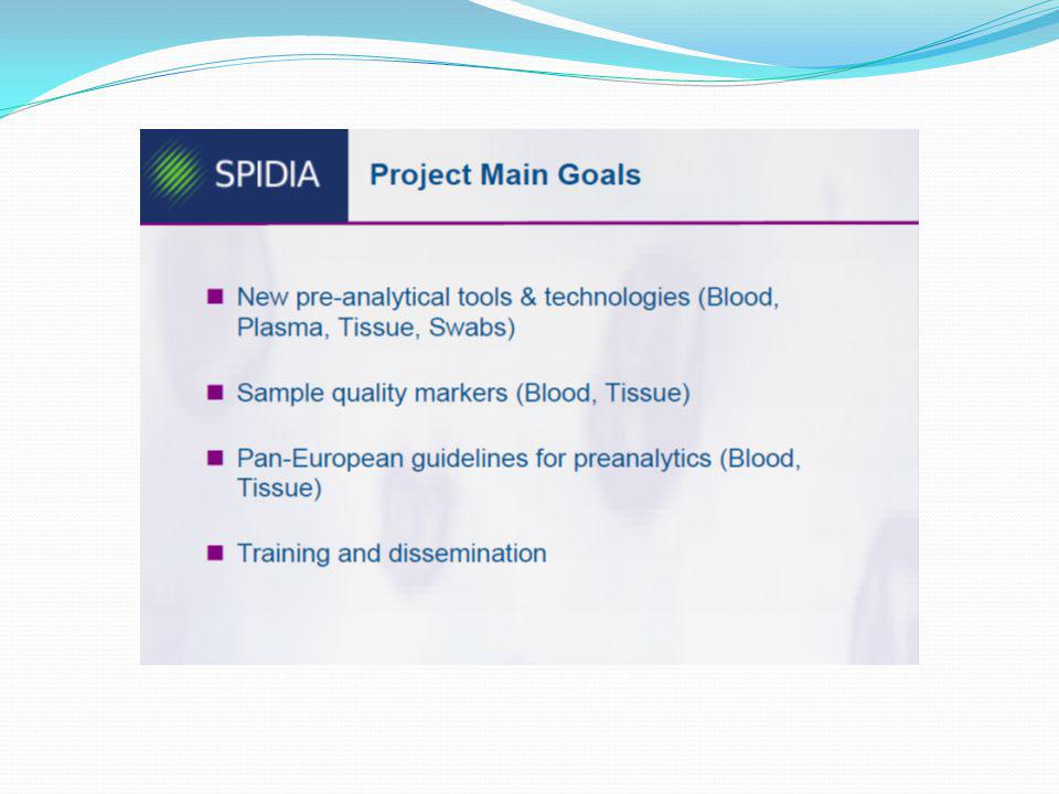
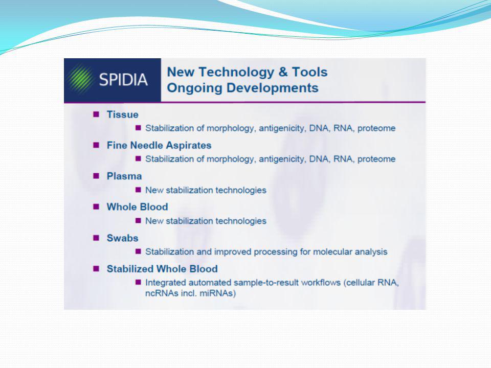
EU SPIDIA PROJESİ Standardization and Improvement of Generic
Pre-analytical Tools and Procedures for In Vitro Diagnostics

36
Sistematik-SPREC-01 Standard Preanalytical Coding for Biospecimens
Each biospecimen is assigned a seven-element-long code that corresponds to seven preanalytical variables and contains a string of 11 (for fluids) or 13 (for solid tissues) letters in a defined order, separated by six hyphens. Wherever possible, we make use of the existing Laboratory Data Management System (LDMS) codes (5) for the sample types and the primary container types. Cancer Epidemiology Biomarkers and Prevention 2010;19:
 or 13 (for solid tissues) letters in a defined order, separated by six hyphens. Wherever possible, we make use of the existing Laboratory Data Management System (LDMS) codes (5) for the sample types and the primary container types. Cancer Epidemiology Biomarkers. and Prevention 2010;19:")
37
Örnekler Sıvı spesmen Serum Plazma İdrar SSS Sample type SER PL2 U24
CSF Type of container SST SED PIX PPT Precentrifugation delay A B Centrifugation E C Second centrifugation N Postcentrifugation delay Storage G J SER-SST-A-E-N-A-G. This corresponds to a serum (SER) specimen that has been collected from a serum collection tube (SST), whose precentrifugation delay is <2 hours at room temperature (A); centrifugation has been done at ambient temperature at 3,000 to 6,000 g with braking (E). Only one centrifugation step was done (N) and the delay between centrifugation and freezing was <1 hour at 3°C to 7°C (A). Serum was stored in straws at a temperature between −85°C and −60°C (G). Plasma specimen PL2-SED-B-B-E-A-G. This corresponds to a double spun plasma (PL2) specimen that has been collected from a sodium EDTA vacutainer collection tube (SED), whose precentrifugation delay is <2 hours at 3°C to 7°C (B); first centrifugation has been done at ambient temperature at <3,000 g with braking (B) and second centrifugation has been done at ambient temperature at 3,000 to 6,000 g with braking (E). The delay between centrifugation and freezing was <1 hour at 3-7°C (A). Plasma was stored in straws at a temperature between −85°C and −60°C (G). Urine specimen U24-PIX-B-A-N-A-J. This corresponds to a 24-hour urine (U24) specimen that has been collected in a collection tube with protease inhibitors (PIX), whose precentrifugation delay is <2 hours at 3°C to 7°C (B); centrifugation has been done at ambient temperature at <3,000 g without braking (A). Only one centrifugation step was done (N) and the delay between centrifugation and freezing was <1 hour at 3°C to 7°C (A). Urine was stored in >5-mL polypropylene tubes at a temperature between −85°C and −60°C (J). CSF Specimen CSF-PPS-B-C-N-A-A. This corresponds to a cerebrospinal fluid (CSF) specimen that has been collected in a sterile polypropylene collection tube (PPS), whose precentrifugation delay is <2 hours at 3°C to 7°C (B); centrifugation has been done at 3°C to 7°C at <3,000 g without braking (C). Only one centrifugation step was done (N) and the delay between centrifugation and freezing was <1 hour at 3°C to 7°C (A). CSF was stored in 0.5- to 2-mL polypropylene tubes at a temperature between −85°C and −60°C (A).
 specimen that has been collected from a serum collection tube (SST), whose precentrifugation delay is <2 hours at room temperature (A); centrifugation has been done at ambient temperature at 3,000 to 6,000 g with braking (E). Only one centrifugation step was done (N) and the delay between centrifugation and freezing was <1 hour at 3°C to 7°C (A). Serum was stored in straws at a temperature between −85°C and −60°C (G). Plasma specimen. PL2-SED-B-B-E-A-G. This corresponds to a double spun plasma (PL2) specimen that has been collected from a sodium EDTA vacutainer collection tube (SED), whose precentrifugation delay is <2 hours at 3°C to 7°C (B); first centrifugation has been done at ambient temperature at <3,000 g with braking (B) and second centrifugation has been done at ambient temperature at 3,000 to 6,000 g with braking (E). The delay between centrifugation and freezing was <1 hour at 3-7°C (A). Plasma was stored in straws at a temperature between −85°C and −60°C (G). Urine specimen. U24-PIX-B-A-N-A-J. This corresponds to a 24-hour urine (U24) specimen that has been collected in a collection tube with protease inhibitors (PIX), whose precentrifugation delay is <2 hours at 3°C to 7°C (B); centrifugation has been done at ambient temperature at <3,000 g without braking (A). Only one centrifugation step was done (N) and the delay between centrifugation and freezing was <1 hour at 3°C to 7°C (A). Urine was stored in >5-mL polypropylene tubes at a temperature between −85°C and −60°C (J). CSF Specimen. CSF-PPS-B-C-N-A-A. This corresponds to a cerebrospinal fluid (CSF) specimen that has been collected in a sterile polypropylene collection tube (PPS), whose precentrifugation delay is <2 hours at 3°C to 7°C (B); centrifugation has been done at 3°C to 7°C at <3,000 g without braking (C). Only one centrifugation step was done (N) and the delay between centrifugation and freezing was <1 hour at 3°C to 7°C (A). CSF was stored in 0.5- to 2-mL polypropylene tubes at a temperature between −85°C and −60°C (A).")
38
Solid spesmen Solid doku Sample type TIS Type of collection BPS
Warm ischemia N Cold ischemia B Fixation type RNL Fixation time A Storage Solid tissue or cytologic Specimen TIS-BPS-N-B-RNL-A-A. This corresponds to a solid tissue (TIS) specimen that has been collected as a biopsy (BPS), with no warm ischemia (N), with cold ischemia of <10 minutes (B), fixed in RNALater (RNL) for <15 minutes (A) and stored in a 0.5- to 2-mL polypropylene tube at a temperature between −85°C and −60°C (A). Biopsies, obtained either at time of traditional surgery, laparoscopy, or puncture, and cytologic specimens such as fine needle aspirates, are assigned the same SPREC. Warm ischemia: Intra-surgical ischemia Cold ischemia: Post-surgical ischemia RNAlater RNA Stabilization Reagent immediately stabilizes RNA in tissue samples to preserve the gene expression profile, and is supplied in 50 ml or 250 ml bottles. For stabilization of DNA, RNA, and protein in tissue samples, Allprotect Tissue Reagent can be used.
 specimen that has been collected as a biopsy (BPS), with no warm ischemia (N), with cold ischemia of <10 minutes (B), fixed in RNALater (RNL) for <15 minutes (A) and stored in a 0.5- to 2-mL polypropylene tube at a temperature between −85°C and −60°C (A). Biopsies, obtained either at time of traditional surgery, laparoscopy, or puncture, and cytologic specimens such as fine needle aspirates, are assigned the same SPREC. Warm ischemia: Intra-surgical ischemia. Cold ischemia: Post-surgical ischemia. RNAlater RNA Stabilization Reagent immediately stabilizes RNA in tissue samples to preserve the gene expression profile, and is supplied in 50 ml or 250 ml bottles. For stabilization of DNA, RNA, and protein in tissue samples, Allprotect Tissue Reagent can be used.")
39
Problemler

40
FFPE ve saklama koşulları
FFPE, Formalin-Fixed, Paraffin-Embedded (tissue). Assessment of protein immunoreactivity to glyceraldehyde-3-phosphate dehydrogenase (GAPDH) by Western blotting. (a) Western Blot by anti-GAPDH antibody. Lane 1, RT-Vac + Drierite; lane 2, RT-HC; lane 3, 4C-Vac + Drierite; lane 4, 4C–HC; lane 5, 30C-Vac + Drierite; lane 6, 30C–HC; lane 7, 37C-Vac + Drierite; lane 8, 37C–HC. (b) Quantitative analysis of Western blot (ImageQuant Program version 5.2, Piscataway, NJ). HC, humidity chamber; RT, room temperature; Vac, vacuum packed. We hypothesize that the presence of both endogenous water and ambient water is crucial to protein degradation and diminution of immunoreactivity in FFPE tissue sections. In an attempt to test our hypothesis, we examined the impact of inadequate processing in FFPE tissues after tissue processing on antigen degradation. To determine the effect of ambient water on protein integrity and immunoreactivity, two storage conditions, vacuum-pack with desiccant and humidity chamber at different temperatures, were further investigated. J Histochem Cytochem April; 59(4): 356–365.
. Assessment of protein immunoreactivity to glyceraldehyde-3-phosphate dehydrogenase (GAPDH) by Western blotting. (a) Western Blot by anti-GAPDH antibody. Lane 1, RT-Vac + Drierite; lane 2, RT-HC; lane 3, 4C-Vac + Drierite; lane 4, 4C–HC; lane 5, 30C-Vac + Drierite; lane 6, 30C–HC; lane 7, 37C-Vac + Drierite; lane 8, 37C–HC. (b) Quantitative analysis of Western blot (ImageQuant Program version 5.2, Piscataway, NJ). HC, humidity chamber; RT, room temperature; Vac, vacuum packed. We hypothesize that the presence of both endogenous water and ambient water is crucial to protein degradation and diminution of immunoreactivity in FFPE tissue sections. In an attempt to test our hypothesis, we examined the impact of inadequate processing in FFPE tissues after tissue processing on antigen degradation. To determine the effect of ambient water on protein integrity and immunoreactivity, two storage conditions, vacuum-pack with desiccant and humidity chamber at different temperatures, were further investigated. J Histochem Cytochem April; 59(4): 356–365.")
41
Warm iskemi ve gen ekspresyonu
WIMA: Warm ischemia-induced metabolic activity WIRD: Warm ischemia-induced RNA degradation Contribution of WIMA and WIRD to changes in gene expression within different time periods. Yi Ma , et al., Analytical Biochemistry Volume 423, Issue

42
Cold iskemi ve gen ekspresyonu
Genlerin yaklaşık % 20-25’i ilk 30 dakikada etkilenmiştir (Affymetrix cDNA microarray) Indivumed, Prof. Dr. Hartmut Juhl, 2nd Biospecimen Research Network Symposium March 16-18, 2009 Kolon kanserinde Affymetrix cDNA microarray sonuçları Following tumor resection ~ 20-25% of genes are differentially expressed within the first 30 minutes ! Sprüssel et al, BioTechniques 2004
 Indivumed, Prof. Dr. Hartmut Juhl, 2nd Biospecimen Research Network Symposium. March 16-18, Kolon kanserinde Affymetrix cDNA microarray sonuçları. Following tumor resection ~ 20-25% of genes are differentially expressed. within the first 30 minutes ! Sprüssel et al, BioTechniques")
43
Cold iskemi ve protein ekspresyonu
Proteinlerin yaklaşık %25-30’u ilk 30 dakikada etkilenmiştir (SELDI-TOF) Tissue ischemia time and protein expression in colon tissue (SELDI-TOF-MS analysis) Following tumor resection ~ 25-30% of proteins are differentially expressed within the first 30 minutes !
 Tissue ischemia time and protein expression in colon tissue (SELDI-TOF-MS analysis) Following tumor resection ~ 25-30% of proteins are differentially expressed. within the first 30 minutes !")
44
Cold iskemi ve IHC/FISH sonuçları
HER2 (Human Epidermal Growth Factor Receptor 2) also known as Neu, ErbB-2, CD340 (cluster of differentiation 340) or p185 is a protein that in humans is encoded by the ERBB2 gene. HER2 is a member of the epidermal growth factor receptor (EGFR/ErbB) family. Amplification or over-expression of this gene has been shown to play an important role in the pathogenesis and progression of certain aggressive types of breast cancer and in recent years it has evolved to become an important biomarker and target of therapy for approx. 30% of breast cancer patients[
 also known as Neu, ErbB-2, CD340 (cluster of differentiation 340) or p185 is a protein that in humans is encoded by the ERBB2 gene. HER2 is a member of the epidermal growth factor receptor (EGFR/ErbB) family. Amplification or over-expression of this gene has been shown to play an important role in the pathogenesis and progression of certain aggressive types of breast cancer and in recent years it has evolved to become an important biomarker and target of therapy for approx. 30% of breast cancer patients[")
45
Biyopsi lokasyonu ve kolon kanserde protein ekspresyonu
Proteinlerin yaklaşık %40’ı tümör bölgesine göre değişim göstermiştir

46
Biyopsi lokasyonu ve kolon kanserde VEGF ekspresyonu
Farklı dokular: 61, 157, 161, 197, 249

47
Sonuç ve Öneriler Sorumluluk tamamen klinisyenlere bırakılamaz;
Sorumluluk büyük oranda laboratuvar uzmanına aittir; Laboratuvar uzmanı, klinisyenleri doğru yönlendirmeli, Sürekli gelişim esas olmalı,

48
Klavuzlar takip edilmeli ve uygulanmalı, Literatür takip edilmeli,
Kalite kontrol sistemleri uygulanmalı, Preanalitik hatalar yanlış teşhis, tedavi ve öngörüye yol açar, Sonuçta hastanın yaşam kalitesini olumsuz etkiler, Biospesmen bilimi’nin varlıgını kabul etmeliyiz What is biospecimen science? Biospecimen Science is the multidisciplinary field of study responsible for establishing tested and proven biospecimen resource-related procedures based on experimentation in the areas of specimen collection, processing, shipping, and storage Why is it needed? Biospecimens are composed of active and reactive living cells or cell products, making them highly complex. The collection, handling, and storage process can profoundly alter the molecular profile and quality of biospecimens. Such alterations, though artificial, can be misinterpreted as disease related or disease specific. High degrees of sensitivity and specificity in new molecular techniques raise the bar for analyte (specimen) data and quality.
 data and quality.")
49
Teşekkürler

50
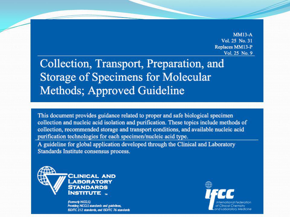
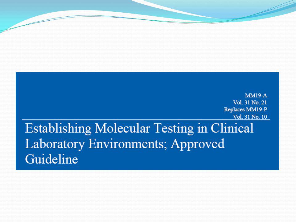
Kılavuzlar (CLSI)
")
Benzer bir sunumlar


>")


>")