Sunuyu indir
Sunum yükleniyor. Lütfen bekleyiniz

1
fetal İskelet dİsplazİlerİ
DOÇ. DR. FİLİZ ÇAYAN MERSİN ÜNİVERSİTESİ TIP FAKÜLTESİ KADIN HASTALIKLARI VE DOĞUM A.D.

2
% 32’si ilk 1 haftada kaybedilir
İSkelet DİSPLAZİLERİ İskeletin yapı, şekil ve dansite anomalilerine neden olan heterojen hastalıklar grubu 2.4/ canlı doğum % 23’ü intrauterin % 32’si ilk 1 haftada kaybedilir

3
PATOLOJİ Genetik geçiş/ yeni mutasyonlar çoğunlukta
350 den fazla farklı klinik tip Ancak 40 tanesine doğumda tanı konabilir

4
Patolojİ EKSTREMİTE DEFEKTLERİ EKSTREMİTE KISALIĞI
ameli : total ekstremite yokluğu meromeli : parsiyel ekstremite yokluğu EKSTREMİTE KISALIĞI Micromelia: ekstremitede total kısalık Rhizomeli: proksimal segmentde Mesomeli: orta segmentde Acromeli: distal segmentde

5
TANI Doğru tanı ? / letalite ?
Görüntüleme yöntemlerinde ki gelişmelere rağmen iskelet displazilerinin intrauterin tanısı güçtür Genellikle kesin tanı ancak yenidoğanın pediatrik veya patolojik incelemesi sonrası konulabilir

6
hafif kozmetik problemlerden letal sonuçlara kadar uzanır
prognoz Çok değişken hafif kozmetik problemlerden letal sonuçlara kadar uzanır

7
European Society of Pediatric Radiology (1977)
sINIFLAMA Geçen 30 yılda klinik, radyolojik ve patolojik sınıflama genetik ve moleküler anormallikleri de içerir hale geldi European Society of Pediatric Radiology (1977) konstitusyonel intrinsik kemik hastalıkları sınıflaması 1983,1997 ve 2001 de modifiye edildi
 konstitusyonel intrinsik kemik hastalıkları sınıflaması 1983,1997 ve 2001 de modifiye edildi.")
8
TanIda zorluklar Farklı tipte anomalilerle birliktelik
Bulguların ortaya çıktığı haftalar değişken Birçoğunda moleküler tanı konulamaz Sistematik değerlendirmede yetersizilik US tek başına yeterli olmayabilir Ekip çalışması ŞART

9
Olguların çoğu OD/OR geçiş gösterdiği için; aile hikayesi pozitif ve fenotipik özellikler tanımlı ise prenatal tanı daha kolay ANCAK tanı genellikle rutin US incelemesi sırasında uzun kemiklerde kısalık veya anormal gelişim saptanması ile şüphelenilerek konulur

10
US US primer tanı yöntemi Sistematik inceleme; Kranyum Toraks Vertebra
Pelvis Uzun kemikler El ve ayaklar

11
İskelet DİSPLAZİSİ DÜŞÜNDÜREN BULGULAR
Kemiklerde; kısalık/ ossifikasyon/ şekil bozuklukları, kırıklar Eklem kontraktürleri Ek bulgular ayırıcı tanıda değerli cloverleaf skull, trident hand, telephone receiver femur, FL/HC < 3 SD FL/AC < 0.16 FL/Foot < 0.8 TC/AC < 0.8 TC/HC < 0.8

12
Worksheet used in the Department of Radiology and Obstetrics at the University of Washington Medical Center while imaging a fetus with suspected skeletal dysplasia Figure 1. Worksheet used in the Department of Radiology and Obstetrics at the University of Washington Medical Center while imaging a fetus with suspected skeletal dysplasia. Dighe M et al. Radiographics 2008;28: ©2008 by Radiological Society of North America

13
Diagram illustrates a diagnostic algorithm for use in fetuses with suspected skeletal dysplasia and skull abnormalities. Figure 8. Diagram illustrates a diagnostic algorithm for use in fetuses with suspected skeletal dysplasia and skull abnormalities. OI = osteogenesis imperfecta. Dighe M et al. Radiographics 2008;28: ©2008 by Radiological Society of North America

14
Diagram illustrates a diagnostic algorithm for use in fetuses with suspected skeletal dysplasia and facial abnormalities. Figure 7. Diagram illustrates a diagnostic algorithm for use in fetuses with suspected skeletal dysplasia and facial abnormalities. OI = osteogenesis imperfecta. Dighe M et al. Radiographics 2008;28: ©2008 by Radiological Society of North America

15
Diagram illustrates a diagnostic algorithm for use in fetuses with suspected skeletal dysplasia and thoracic abnormalities. Figure 6. Diagram illustrates a diagnostic algorithm for use in fetuses with suspected skeletal dysplasia and thoracic abnormalities. OI = osteogenesis imperfecta. Dighe M et al. Radiographics 2008;28: ©2008 by Radiological Society of North America

16
İskelet displazi olasılığı yüksek
KISA FEMUR FL < 2 SD Normal fizyolojik varyasyon IUGR Fokal kısalık Aneuploidi *Trizomi 21 fetuslarda , normal fetuslara oranla kısa femur x 4 kat fazla görülür FL < 2 SD – 5mm İskelet displazi olasılığı yüksek

17
TORAKS Toraks çevresi ve kardio-torasik oran
4 kadran görüntüsü düzeyinde ölçülür < 5 percentile pulmoner hipoplazi Uzun ve dar thorax Jeune sendromu Kosta kırıkları osteogenesis imperfekta Kostalar ileri derecede kısa short rib polidaktili sendromu Klavikula (-) cleidokranial displazi Scapula (-) camptomelic displazi
 cleidokranial displazi. Scapula (-) camptomelic displazi.")
18
Preaxial /postaxial polidaktili
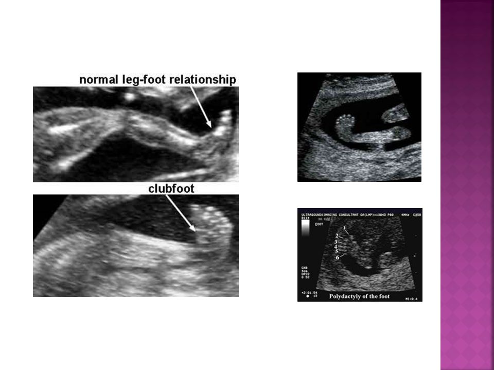
EL VE AYAKLAR Preaxial /postaxial polidaktili Sindaktili Klinodaktili Hipoplazi /aplaziler Postural deformiteler: hitchhiker’s thumb,rocker-bottom feet, clubbed feet, hand, clenched hand

20
US image shows a typical clenched hand of a fetus with trisomy 18 at 18 weeks of gestation.
Figure 4b. (a) US image shows a typical clenched hand of a fetus with trisomy 18 at 18 weeks of gestation. (b) US image shows ulnar deviation of the index finger, which overlaps the other digits. Rypens F et al. Radiographics 2006;26: ©2006 by Radiological Society of North America
 US image shows a typical clenched hand of a fetus with trisomy 18 at 18 weeks of gestation. (b) US image shows ulnar deviation of the index finger, which overlaps the other digits. Rypens F et al. Radiographics 2006;26: ©2006 by Radiological Society of North America.")
21
Focal akinesia Figure 5a. Focal akinesia. (a) Schematic representation of the anomalies observed at US in a fetus at 30 weeks of gestation. (b) US image shows camptodactyly, with permanent flexion of the proximal phalanges of the third and fourth fingers (arrows) and with flexion deformities of the distal interphalangeal articulations of the second and fifth fingers, which are extended. (c) US image, which shows the palmar aspect of the same hand, helps confirm the proximal flexion of the third and fourth fingers (arrows). Rypens F et al. Radiographics 2006;26: ©2006 by Radiological Society of North America
 Schematic representation of the anomalies observed at US in a fetus at 30 weeks of gestation. (b) US image shows camptodactyly, with permanent flexion of the proximal phalanges of the third and fourth fingers (arrows) and with flexion deformities of the distal interphalangeal articulations of the second and fifth fingers, which are extended. (c) US image, which shows the palmar aspect of the same hand, helps confirm the proximal flexion of the third and fourth fingers (arrows). Rypens F et al. Radiographics 2006;26: ©2006 by Radiological Society of North America.")
22
Diffuse akinesia. Figure 7b. Diffuse akinesia. (a) US image in a fetus at 23 weeks of gestation shows the left wrist flexed, the hand closed, and the elbow extended, a position that remained unchanged during two successive examinations. (b) Corresponding radiograph of the left upper limb of the same fetus shows a clubhand and a normal radius and ulna. The other three limbs (not shown) also manifested fixed deformities. Rypens F et al. Radiographics 2006;26: ©2006 by Radiological Society of North America
 US image in a fetus at 23 weeks of gestation shows the left wrist flexed, the hand closed, and the elbow extended, a position that remained unchanged during two successive examinations. (b) Corresponding radiograph of the left upper limb of the same fetus shows a clubhand and a normal radius and ulna. The other three limbs (not shown) also manifested fixed deformities. Rypens F et al. Radiographics 2006;26: ©2006 by Radiological Society of North America.")
23
Phocomelia Figure 10a. Phocomelia. (a) Coronal oblique US image of a fetal thorax at 20 weeks of gestation shows abnormal direct implantation of the hand (arrow) on the thorax. (b) Corresponding radiograph shows the bilateral anomaly. Rypens F et al. Radiographics 2006;26: ©2006 by Radiological Society of North America
 Coronal oblique US image of a fetal thorax at 20 weeks of gestation shows abnormal direct implantation of the hand (arrow) on the thorax. (b) Corresponding radiograph shows the bilateral anomaly. Rypens F et al. Radiographics 2006;26: ©2006 by Radiological Society of North America.")
24
KRANİUM Şekil, biyometri ve mineralizasyon değerlendirilir
Makrosefali, mikrosefali frontal bossing, cloverleaf skull hiper-, hipotelorizm Mikrognati Kulak anomalileri Şekil anomalileri Brakisefali Dolikosefali Skafosefali Kraniosinositoz

25
Kırıklar, mineralizasyon
VERTEBRA Nöral tüp defekti Skolyoz Platispondili Kırıklar, mineralizasyon

26
Arnold-Chiari malformation with the banana sign and meningomyelocele at 14 weeks gestation
Figure 7b. Arnold-Chiari malformation with the banana sign and meningomyelocele at 14 weeks gestation. (a) Axial transabdominal US image of the fetal head shows a banana-shaped cerebellum (arrows) and effacement of the cisterna magna (CM). (b) Sagittal US image of the fetal lumbosacral region shows a meningomyelocele (arrow). Fong K W et al. Radiographics 2004;24: ©2004 by Radiological Society of North America
 Axial transabdominal US image of the fetal head shows a banana-shaped cerebellum (arrows) and effacement of the cisterna magna (CM). (b) Sagittal US image of the fetal lumbosacral region shows a meningomyelocele (arrow). Fong K W et al. Radiographics 2004;24: ©2004 by Radiological Society of North America.")
27
İskelet DİSPLAZİLERİ ÖRNEKLERİ

28
EKSTREMİTE YOKLUĞU 0.5/ 10.000 Genellikle izole
Amniotik band, teratojen, vaskuler kazalar Amelia Meromelia

29
Isolated limb deficiency
Figure 9a. Isolated limb deficiency. (a, b) Two-dimensional (a) and 3D (b) US images show a shortened forearm with an abnormal hand (arrow) (note the lack of a normal hand and the abnormal soft tissue at the distal end of the forearm), normal limb bone echogenicity, and otherwise normal anatomy. According to the diagram in Figure 4, the patient appeared to have distal limb agenesis as an isolated finding. (c) Radiograph shows abnormal bone tissue (arrow) at the end of the normally formed and mineralized forearm bone. (d) Autopsy photograph shows an abnormal left hand with five tiny fingers and apparent fingernails (arrow). Dighe M et al. Radiographics 2008;28: ©2008 by Radiological Society of North America
 Two-dimensional (a) and 3D (b) US images show a shortened forearm with an abnormal hand (arrow) (note the lack of a normal hand and the abnormal soft tissue at the distal end of the forearm), normal limb bone echogenicity, and otherwise normal anatomy. According to the diagram in Figure 4, the patient appeared to have distal limb agenesis as an isolated finding. (c) Radiograph shows abnormal bone tissue (arrow) at the end of the normally formed and mineralized forearm bone. (d) Autopsy photograph shows an abnormal left hand with five tiny fingers and apparent fingernails (arrow). Dighe M et al. Radiographics 2008;28: ©2008 by Radiological Society of North America.")
30
Isolated limb deficiency
Figure 9b. Isolated limb deficiency. (a, b) Two-dimensional (a) and 3D (b) US images show a shortened forearm with an abnormal hand (arrow) (note the lack of a normal hand and the abnormal soft tissue at the distal end of the forearm), normal limb bone echogenicity, and otherwise normal anatomy. According to the diagram in Figure 4, the patient appeared to have distal limb agenesis as an isolated finding. (c) Radiograph shows abnormal bone tissue (arrow) at the end of the normally formed and mineralized forearm bone. (d) Autopsy photograph shows an abnormal left hand with five tiny fingers and apparent fingernails (arrow). Dighe M et al. Radiographics 2008;28: ©2008 by Radiological Society of North America
 Two-dimensional (a) and 3D (b) US images show a shortened forearm with an abnormal hand (arrow) (note the lack of a normal hand and the abnormal soft tissue at the distal end of the forearm), normal limb bone echogenicity, and otherwise normal anatomy. According to the diagram in Figure 4, the patient appeared to have distal limb agenesis as an isolated finding. (c) Radiograph shows abnormal bone tissue (arrow) at the end of the normally formed and mineralized forearm bone. (d) Autopsy photograph shows an abnormal left hand with five tiny fingers and apparent fingernails (arrow). Dighe M et al. Radiographics 2008;28: ©2008 by Radiological Society of North America.")
31
Isolated limb deficiency
Figure 9d. Isolated limb deficiency. (a, b) Two-dimensional (a) and 3D (b) US images show a shortened forearm with an abnormal hand (arrow) (note the lack of a normal hand and the abnormal soft tissue at the distal end of the forearm), normal limb bone echogenicity, and otherwise normal anatomy. According to the diagram in Figure 4, the patient appeared to have distal limb agenesis as an isolated finding. (c) Radiograph shows abnormal bone tissue (arrow) at the end of the normally formed and mineralized forearm bone. (d) Autopsy photograph shows an abnormal left hand with five tiny fingers and apparent fingernails (arrow). Dighe M et al. Radiographics 2008;28: ©2008 by Radiological Society of North America
 Two-dimensional (a) and 3D (b) US images show a shortened forearm with an abnormal hand (arrow) (note the lack of a normal hand and the abnormal soft tissue at the distal end of the forearm), normal limb bone echogenicity, and otherwise normal anatomy. According to the diagram in Figure 4, the patient appeared to have distal limb agenesis as an isolated finding. (c) Radiograph shows abnormal bone tissue (arrow) at the end of the normally formed and mineralized forearm bone. (d) Autopsy photograph shows an abnormal left hand with five tiny fingers and apparent fingernails (arrow). Dighe M et al. Radiographics 2008;28: ©2008 by Radiological Society of North America.")
32
TANATROFİK DİSPLAZİ Tanatrofik « ölümcül »
En sık letal fetal iskelet displazisi Fibroblast growth factor receptor 3 (FGFR3) gen mutasyonu OD (genellikle) 2 tipi var Genellikle erken yenidoğan döneminde pulmoner hipoplazi nedeniyle kaybedilir
 gen mutasyonu. OD (genellikle) 2 tipi var. Genellikle erken yenidoğan döneminde pulmoner hipoplazi nedeniyle kaybedilir.")
33
Tip 1 Tip 2 Ciddi ekstremite kısalığı Tip 1’ de eğimli Tip 2’ de düz
makrosefali cloverleaf frontal bossing burun kökü basık trident hand torakal hipoplazi telephone receiver Ekstremitelerde ileri derecede açılanma ve kısalık Uzun kemikler düz ve kısa

34
Thanatophoric dysplasia type 1
Figure 10a. Thanatophoric dysplasia type 1. (a) Transverse US image shows a normal-shaped but enlarged head. (b) Sagittal US image shows a depressed nasal bridge (arrowhead), a prominent forehead (double arrows), and an undersized thorax (single arrow) compared with the abdomen. (c) US image shows a telephone receiver–shaped femur (arrows). Normal limb echogenicity with severe shortening and bowing of the limbs, a narrow chest, and macrocephaly suggest thanatophoric dysplasia type 1 according to the diagrams in Figures 2, 6, and 8, respectively. (d) Postmortem radiograph shows bowed long bones (white arrows), a narrow chest, and platyspondyly (black arrow). (e, f) Autopsy photographs show shortened limbs, the depressed nasal bridge (arrowhead in f), a short trunk, an enlarged abdomen, and the prominent forehead (arrows in f). Dighe M et al. Radiographics 2008;28: ©2008 by Radiological Society of North America
 Transverse US image shows a normal-shaped but enlarged head. (b) Sagittal US image shows a depressed nasal bridge (arrowhead), a prominent forehead (double arrows), and an undersized thorax (single arrow) compared with the abdomen. (c) US image shows a telephone receiver–shaped femur (arrows). Normal limb echogenicity with severe shortening and bowing of the limbs, a narrow chest, and macrocephaly suggest thanatophoric dysplasia type 1 according to the diagrams in Figures 2, 6, and 8, respectively. (d) Postmortem radiograph shows bowed long bones (white arrows), a narrow chest, and platyspondyly (black arrow). (e, f) Autopsy photographs show shortened limbs, the depressed nasal bridge (arrowhead in f), a short trunk, an enlarged abdomen, and the prominent forehead (arrows in f). Dighe M et al. Radiographics 2008;28: ©2008 by Radiological Society of North America.")
35
Thanatophoric dysplasia type 1
Figure 10b. Thanatophoric dysplasia type 1. (a) Transverse US image shows a normal-shaped but enlarged head. (b) Sagittal US image shows a depressed nasal bridge (arrowhead), a prominent forehead (double arrows), and an undersized thorax (single arrow) compared with the abdomen. (c) US image shows a telephone receiver–shaped femur (arrows). Normal limb echogenicity with severe shortening and bowing of the limbs, a narrow chest, and macrocephaly suggest thanatophoric dysplasia type 1 according to the diagrams in Figures 2, 6, and 8, respectively. (d) Postmortem radiograph shows bowed long bones (white arrows), a narrow chest, and platyspondyly (black arrow). (e, f) Autopsy photographs show shortened limbs, the depressed nasal bridge (arrowhead in f), a short trunk, an enlarged abdomen, and the prominent forehead (arrows in f). Dighe M et al. Radiographics 2008;28: ©2008 by Radiological Society of North America
 Transverse US image shows a normal-shaped but enlarged head. (b) Sagittal US image shows a depressed nasal bridge (arrowhead), a prominent forehead (double arrows), and an undersized thorax (single arrow) compared with the abdomen. (c) US image shows a telephone receiver–shaped femur (arrows). Normal limb echogenicity with severe shortening and bowing of the limbs, a narrow chest, and macrocephaly suggest thanatophoric dysplasia type 1 according to the diagrams in Figures 2, 6, and 8, respectively. (d) Postmortem radiograph shows bowed long bones (white arrows), a narrow chest, and platyspondyly (black arrow). (e, f) Autopsy photographs show shortened limbs, the depressed nasal bridge (arrowhead in f), a short trunk, an enlarged abdomen, and the prominent forehead (arrows in f). Dighe M et al. Radiographics 2008;28: ©2008 by Radiological Society of North America.")
36
Thanatophoric dysplasia type 1
Figure 10c. Thanatophoric dysplasia type 1. (a) Transverse US image shows a normal-shaped but enlarged head. (b) Sagittal US image shows a depressed nasal bridge (arrowhead), a prominent forehead (double arrows), and an undersized thorax (single arrow) compared with the abdomen. (c) US image shows a telephone receiver–shaped femur (arrows). Normal limb echogenicity with severe shortening and bowing of the limbs, a narrow chest, and macrocephaly suggest thanatophoric dysplasia type 1 according to the diagrams in Figures 2, 6, and 8, respectively. (d) Postmortem radiograph shows bowed long bones (white arrows), a narrow chest, and platyspondyly (black arrow). (e, f) Autopsy photographs show shortened limbs, the depressed nasal bridge (arrowhead in f), a short trunk, an enlarged abdomen, and the prominent forehead (arrows in f). Dighe M et al. Radiographics 2008;28: ©2008 by Radiological Society of North America
 Transverse US image shows a normal-shaped but enlarged head. (b) Sagittal US image shows a depressed nasal bridge (arrowhead), a prominent forehead (double arrows), and an undersized thorax (single arrow) compared with the abdomen. (c) US image shows a telephone receiver–shaped femur (arrows). Normal limb echogenicity with severe shortening and bowing of the limbs, a narrow chest, and macrocephaly suggest thanatophoric dysplasia type 1 according to the diagrams in Figures 2, 6, and 8, respectively. (d) Postmortem radiograph shows bowed long bones (white arrows), a narrow chest, and platyspondyly (black arrow). (e, f) Autopsy photographs show shortened limbs, the depressed nasal bridge (arrowhead in f), a short trunk, an enlarged abdomen, and the prominent forehead (arrows in f). Dighe M et al. Radiographics 2008;28: ©2008 by Radiological Society of North America.")
37
Thanatophoric dysplasia type 1
Figure 10d. Thanatophoric dysplasia type 1. (a) Transverse US image shows a normal-shaped but enlarged head. (b) Sagittal US image shows a depressed nasal bridge (arrowhead), a prominent forehead (double arrows), and an undersized thorax (single arrow) compared with the abdomen. (c) US image shows a telephone receiver–shaped femur (arrows). Normal limb echogenicity with severe shortening and bowing of the limbs, a narrow chest, and macrocephaly suggest thanatophoric dysplasia type 1 according to the diagrams in Figures 2, 6, and 8, respectively. (d) Postmortem radiograph shows bowed long bones (white arrows), a narrow chest, and platyspondyly (black arrow). (e, f) Autopsy photographs show shortened limbs, the depressed nasal bridge (arrowhead in f), a short trunk, an enlarged abdomen, and the prominent forehead (arrows in f). Dighe M et al. Radiographics 2008;28: ©2008 by Radiological Society of North America
 Transverse US image shows a normal-shaped but enlarged head. (b) Sagittal US image shows a depressed nasal bridge (arrowhead), a prominent forehead (double arrows), and an undersized thorax (single arrow) compared with the abdomen. (c) US image shows a telephone receiver–shaped femur (arrows). Normal limb echogenicity with severe shortening and bowing of the limbs, a narrow chest, and macrocephaly suggest thanatophoric dysplasia type 1 according to the diagrams in Figures 2, 6, and 8, respectively. (d) Postmortem radiograph shows bowed long bones (white arrows), a narrow chest, and platyspondyly (black arrow). (e, f) Autopsy photographs show shortened limbs, the depressed nasal bridge (arrowhead in f), a short trunk, an enlarged abdomen, and the prominent forehead (arrows in f). Dighe M et al. Radiographics 2008;28: ©2008 by Radiological Society of North America.")
38
Thanatophoric dysplasia type 1
Figure 10f. Thanatophoric dysplasia type 1. (a) Transverse US image shows a normal-shaped but enlarged head. (b) Sagittal US image shows a depressed nasal bridge (arrowhead), a prominent forehead (double arrows), and an undersized thorax (single arrow) compared with the abdomen. (c) US image shows a telephone receiver–shaped femur (arrows). Normal limb echogenicity with severe shortening and bowing of the limbs, a narrow chest, and macrocephaly suggest thanatophoric dysplasia type 1 according to the diagrams in Figures 2, 6, and 8, respectively. (d) Postmortem radiograph shows bowed long bones (white arrows), a narrow chest, and platyspondyly (black arrow). (e, f) Autopsy photographs show shortened limbs, the depressed nasal bridge (arrowhead in f), a short trunk, an enlarged abdomen, and the prominent forehead (arrows in f). Dighe M et al. Radiographics 2008;28: ©2008 by Radiological Society of North America
 Transverse US image shows a normal-shaped but enlarged head. (b) Sagittal US image shows a depressed nasal bridge (arrowhead), a prominent forehead (double arrows), and an undersized thorax (single arrow) compared with the abdomen. (c) US image shows a telephone receiver–shaped femur (arrows). Normal limb echogenicity with severe shortening and bowing of the limbs, a narrow chest, and macrocephaly suggest thanatophoric dysplasia type 1 according to the diagrams in Figures 2, 6, and 8, respectively. (d) Postmortem radiograph shows bowed long bones (white arrows), a narrow chest, and platyspondyly (black arrow). (e, f) Autopsy photographs show shortened limbs, the depressed nasal bridge (arrowhead in f), a short trunk, an enlarged abdomen, and the prominent forehead (arrows in f). Dighe M et al. Radiographics 2008;28: ©2008 by Radiological Society of North America.")
39
Thanatophoric dysplasia type 2
Figure 11a. Thanatophoric dysplasia type 2. (a) Axial US image shows an oversized head with a cloverleaf shape (arrows). (b) Sagittal US image shows a temporal bulge (arrow). (c) US image shows a “trident” hand. Normal limb echogenicity with severe shortening, a narrow chest, and an irregular shape of the head suggest thanatophoric dysplasia type 2 according to the diagrams in Figures 2, 6, and 8, respectively. (d) US image shows a short but relatively straight long bone. (e) Coronal US image through the abdomen-chest shows a hypoplastic thorax (arrow). (f) Radiograph shows the cloverleaf skull shape created by the temporal bulge in the skull (arrow). (g, h) Postmortem photographs show the prominent forehead; the typical temporal bulge, resulting in the cloverleaf skull shape (double arrows); and the trident hand (single arrow in h). Note the bulge in the occipital region, a finding that represents an occipital encephalocele (an unusual finding in thanatophoric dysplasia). Dighe M et al. Radiographics 2008;28: ©2008 by Radiological Society of North America
 Axial US image shows an oversized head with a cloverleaf shape (arrows). (b) Sagittal US image shows a temporal bulge (arrow). (c) US image shows a trident hand. Normal limb echogenicity with severe shortening, a narrow chest, and an irregular shape of the head suggest thanatophoric dysplasia type 2 according to the diagrams in Figures 2, 6, and 8, respectively. (d) US image shows a short but relatively straight long bone. (e) Coronal US image through the abdomen-chest shows a hypoplastic thorax (arrow). (f) Radiograph shows the cloverleaf skull shape created by the temporal bulge in the skull (arrow). (g, h) Postmortem photographs show the prominent forehead; the typical temporal bulge, resulting in the cloverleaf skull shape (double arrows); and the trident hand (single arrow in h). Note the bulge in the occipital region, a finding that represents an occipital encephalocele (an unusual finding in thanatophoric dysplasia). Dighe M et al. Radiographics 2008;28: ©2008 by Radiological Society of North America.")
40
Thanatophoric dysplasia type 2
Figure 11c. Thanatophoric dysplasia type 2. (a) Axial US image shows an oversized head with a cloverleaf shape (arrows). (b) Sagittal US image shows a temporal bulge (arrow). (c) US image shows a “trident” hand. Normal limb echogenicity with severe shortening, a narrow chest, and an irregular shape of the head suggest thanatophoric dysplasia type 2 according to the diagrams in Figures 2, 6, and 8, respectively. (d) US image shows a short but relatively straight long bone. (e) Coronal US image through the abdomen-chest shows a hypoplastic thorax (arrow). (f) Radiograph shows the cloverleaf skull shape created by the temporal bulge in the skull (arrow). (g, h) Postmortem photographs show the prominent forehead; the typical temporal bulge, resulting in the cloverleaf skull shape (double arrows); and the trident hand (single arrow in h). Note the bulge in the occipital region, a finding that represents an occipital encephalocele (an unusual finding in thanatophoric dysplasia). Dighe M et al. Radiographics 2008;28: ©2008 by Radiological Society of North America
 Axial US image shows an oversized head with a cloverleaf shape (arrows). (b) Sagittal US image shows a temporal bulge (arrow). (c) US image shows a trident hand. Normal limb echogenicity with severe shortening, a narrow chest, and an irregular shape of the head suggest thanatophoric dysplasia type 2 according to the diagrams in Figures 2, 6, and 8, respectively. (d) US image shows a short but relatively straight long bone. (e) Coronal US image through the abdomen-chest shows a hypoplastic thorax (arrow). (f) Radiograph shows the cloverleaf skull shape created by the temporal bulge in the skull (arrow). (g, h) Postmortem photographs show the prominent forehead; the typical temporal bulge, resulting in the cloverleaf skull shape (double arrows); and the trident hand (single arrow in h). Note the bulge in the occipital region, a finding that represents an occipital encephalocele (an unusual finding in thanatophoric dysplasia). Dighe M et al. Radiographics 2008;28: ©2008 by Radiological Society of North America.")
41
Thanatophoric dysplasia type 2.
Figure 11f. Thanatophoric dysplasia type 2. (a) Axial US image shows an oversized head with a cloverleaf shape (arrows). (b) Sagittal US image shows a temporal bulge (arrow). (c) US image shows a “trident” hand. Normal limb echogenicity with severe shortening, a narrow chest, and an irregular shape of the head suggest thanatophoric dysplasia type 2 according to the diagrams in Figures 2, 6, and 8, respectively. (d) US image shows a short but relatively straight long bone. (e) Coronal US image through the abdomen-chest shows a hypoplastic thorax (arrow). (f) Radiograph shows the cloverleaf skull shape created by the temporal bulge in the skull (arrow). (g, h) Postmortem photographs show the prominent forehead; the typical temporal bulge, resulting in the cloverleaf skull shape (double arrows); and the trident hand (single arrow in h). Note the bulge in the occipital region, a finding that represents an occipital encephalocele (an unusual finding in thanatophoric dysplasia). Dighe M et al. Radiographics 2008;28: ©2008 by Radiological Society of North America
 Axial US image shows an oversized head with a cloverleaf shape (arrows). (b) Sagittal US image shows a temporal bulge (arrow). (c) US image shows a trident hand. Normal limb echogenicity with severe shortening, a narrow chest, and an irregular shape of the head suggest thanatophoric dysplasia type 2 according to the diagrams in Figures 2, 6, and 8, respectively. (d) US image shows a short but relatively straight long bone. (e) Coronal US image through the abdomen-chest shows a hypoplastic thorax (arrow). (f) Radiograph shows the cloverleaf skull shape created by the temporal bulge in the skull (arrow). (g, h) Postmortem photographs show the prominent forehead; the typical temporal bulge, resulting in the cloverleaf skull shape (double arrows); and the trident hand (single arrow in h). Note the bulge in the occipital region, a finding that represents an occipital encephalocele (an unusual finding in thanatophoric dysplasia). Dighe M et al. Radiographics 2008;28: ©2008 by Radiological Society of North America.")
42
Thanatophoric dysplasia type 2
Figure 11g. Thanatophoric dysplasia type 2. (a) Axial US image shows an oversized head with a cloverleaf shape (arrows). (b) Sagittal US image shows a temporal bulge (arrow). (c) US image shows a “trident” hand. Normal limb echogenicity with severe shortening, a narrow chest, and an irregular shape of the head suggest thanatophoric dysplasia type 2 according to the diagrams in Figures 2, 6, and 8, respectively. (d) US image shows a short but relatively straight long bone. (e) Coronal US image through the abdomen-chest shows a hypoplastic thorax (arrow). (f) Radiograph shows the cloverleaf skull shape created by the temporal bulge in the skull (arrow). (g, h) Postmortem photographs show the prominent forehead; the typical temporal bulge, resulting in the cloverleaf skull shape (double arrows); and the trident hand (single arrow in h). Note the bulge in the occipital region, a finding that represents an occipital encephalocele (an unusual finding in thanatophoric dysplasia). Dighe M et al. Radiographics 2008;28: ©2008 by Radiological Society of North America
 Axial US image shows an oversized head with a cloverleaf shape (arrows). (b) Sagittal US image shows a temporal bulge (arrow). (c) US image shows a trident hand. Normal limb echogenicity with severe shortening, a narrow chest, and an irregular shape of the head suggest thanatophoric dysplasia type 2 according to the diagrams in Figures 2, 6, and 8, respectively. (d) US image shows a short but relatively straight long bone. (e) Coronal US image through the abdomen-chest shows a hypoplastic thorax (arrow). (f) Radiograph shows the cloverleaf skull shape created by the temporal bulge in the skull (arrow). (g, h) Postmortem photographs show the prominent forehead; the typical temporal bulge, resulting in the cloverleaf skull shape (double arrows); and the trident hand (single arrow in h). Note the bulge in the occipital region, a finding that represents an occipital encephalocele (an unusual finding in thanatophoric dysplasia). Dighe M et al. Radiographics 2008;28: ©2008 by Radiological Society of North America.")
43
Osteogenezİs İmperfekta
0.4/ canlı doğum Tip 1 kollajen gen defekti nedeni ile kemik frajilitesi artmış Genellikle OD kalıtım gösterir ve yeni mutasyonlarla oluşur

44
Osteogenezİs İmperfekta
Klinik ve genetik olarak heterojen bir grup 4 tipi mevcut (klinik, genetik, radyografik sınıflama) Tip 2; Genellikle inutero veya erken yenidoğan döneminde pulmoner hipoplazi veya ciddi kırıklar nedeni ile ex olur
 Tip 2; Genellikle inutero veya erken yenidoğan döneminde pulmoner hipoplazi veya ciddi kırıklar nedeni ile ex olur.")
45
Osteogenezİs İmperfekta
USG bulguları Ciddi mikromeli (FL< 3 SD) Torakal hipoplazi Gövde kısalığı Normal kranyum Kırıklar ( tek veya birçok ) Azalmış mineralizasyon Konkav torasik kontur Kemiklerde multiple açılanma/ callus Kranyumda US transduser bası deformasyonu
 Torakal hipoplazi. Gövde kısalığı. Normal kranyum. Kırıklar ( tek veya birçok ) Azalmış mineralizasyon. Konkav torasik kontur. Kemiklerde multiple açılanma/ callus. Kranyumda US transduser bası deformasyonu.")
46
Osteogenesis imperfecta.
kırıklar Figure 12a. Osteogenesis imperfecta. (a–c) US images show bone fractures and deformities. Note the femoral irregularity and angulation (arrow in a), a finding that is consistent with fractures; the decreased skull ossification, which allows easy visualization of the intracranial structures (b); and the irregular shape of the ribs (arrow in c), a finding that also suggests fractures. Decreased echogenicity of the limbs with shortening, decreased echogenicity of the ribs with fractures, and hypoechogenicity of the head suggest osteogenesis imperfecta according to the diagrams in Figures 5, 6, and 8, respectively. (d) Postmortem photograph shows irregular ribs (arrow) due to healing fractures. (e) Postmortem photograph shows deformed extremities, findings that are consistent with fractures. (f) Postmortem radiograph shows wavy ribs (black arrow) and irregular deformed long bones (white arrows) due to multiple fractures. Dighe M et al. Radiographics 2008;28: ©2008 by Radiological Society of North America
 US images show bone fractures and deformities. Note the femoral irregularity and angulation (arrow in a), a finding that is consistent with fractures; the decreased skull ossification, which allows easy visualization of the intracranial structures (b); and the irregular shape of the ribs (arrow in c), a finding that also suggests fractures. Decreased echogenicity of the limbs with shortening, decreased echogenicity of the ribs with fractures, and hypoechogenicity of the head suggest osteogenesis imperfecta according to the diagrams in Figures 5, 6, and 8, respectively. (d) Postmortem photograph shows irregular ribs (arrow) due to healing fractures. (e) Postmortem photograph shows deformed extremities, findings that are consistent with fractures. (f) Postmortem radiograph shows wavy ribs (black arrow) and irregular deformed long bones (white arrows) due to multiple fractures. Dighe M et al. Radiographics 2008;28: ©2008 by Radiological Society of North America.")
47
Osteogenesis imperfecta.
Azalmış ossifikasyon transduser basısı Figure 12b. Osteogenesis imperfecta. (a–c) US images show bone fractures and deformities. Note the femoral irregularity and angulation (arrow in a), a finding that is consistent with fractures; the decreased skull ossification, which allows easy visualization of the intracranial structures (b); and the irregular shape of the ribs (arrow in c), a finding that also suggests fractures. Decreased echogenicity of the limbs with shortening, decreased echogenicity of the ribs with fractures, and hypoechogenicity of the head suggest osteogenesis imperfecta according to the diagrams in Figures 5, 6, and 8, respectively. (d) Postmortem photograph shows irregular ribs (arrow) due to healing fractures. (e) Postmortem photograph shows deformed extremities, findings that are consistent with fractures. (f) Postmortem radiograph shows wavy ribs (black arrow) and irregular deformed long bones (white arrows) due to multiple fractures. Dighe M et al. Radiographics 2008;28: ©2008 by Radiological Society of North America
 US images show bone fractures and deformities. Note the femoral irregularity and angulation (arrow in a), a finding that is consistent with fractures; the decreased skull ossification, which allows easy visualization of the intracranial structures (b); and the irregular shape of the ribs (arrow in c), a finding that also suggests fractures. Decreased echogenicity of the limbs with shortening, decreased echogenicity of the ribs with fractures, and hypoechogenicity of the head suggest osteogenesis imperfecta according to the diagrams in Figures 5, 6, and 8, respectively. (d) Postmortem photograph shows irregular ribs (arrow) due to healing fractures. (e) Postmortem photograph shows deformed extremities, findings that are consistent with fractures. (f) Postmortem radiograph shows wavy ribs (black arrow) and irregular deformed long bones (white arrows) due to multiple fractures. Dighe M et al. Radiographics 2008;28: ©2008 by Radiological Society of North America.")
48
Osteogenesis imperfecta.
İrregularite ve kırıklar Figure 12c. Osteogenesis imperfecta. (a–c) US images show bone fractures and deformities. Note the femoral irregularity and angulation (arrow in a), a finding that is consistent with fractures; the decreased skull ossification, which allows easy visualization of the intracranial structures (b); and the irregular shape of the ribs (arrow in c), a finding that also suggests fractures. Decreased echogenicity of the limbs with shortening, decreased echogenicity of the ribs with fractures, and hypoechogenicity of the head suggest osteogenesis imperfecta according to the diagrams in Figures 5, 6, and 8, respectively. (d) Postmortem photograph shows irregular ribs (arrow) due to healing fractures. (e) Postmortem photograph shows deformed extremities, findings that are consistent with fractures. (f) Postmortem radiograph shows wavy ribs (black arrow) and irregular deformed long bones (white arrows) due to multiple fractures. Dighe M et al. Radiographics 2008;28: ©2008 by Radiological Society of North America
 US images show bone fractures and deformities. Note the femoral irregularity and angulation (arrow in a), a finding that is consistent with fractures; the decreased skull ossification, which allows easy visualization of the intracranial structures (b); and the irregular shape of the ribs (arrow in c), a finding that also suggests fractures. Decreased echogenicity of the limbs with shortening, decreased echogenicity of the ribs with fractures, and hypoechogenicity of the head suggest osteogenesis imperfecta according to the diagrams in Figures 5, 6, and 8, respectively. (d) Postmortem photograph shows irregular ribs (arrow) due to healing fractures. (e) Postmortem photograph shows deformed extremities, findings that are consistent with fractures. (f) Postmortem radiograph shows wavy ribs (black arrow) and irregular deformed long bones (white arrows) due to multiple fractures. Dighe M et al. Radiographics 2008;28: ©2008 by Radiological Society of North America.")
49
Osteogenesis imperfecta.
Tekrarlayan kırıklar callus formasyonu Figure 12d. Osteogenesis imperfecta. (a–c) US images show bone fractures and deformities. Note the femoral irregularity and angulation (arrow in a), a finding that is consistent with fractures; the decreased skull ossification, which allows easy visualization of the intracranial structures (b); and the irregular shape of the ribs (arrow in c), a finding that also suggests fractures. Decreased echogenicity of the limbs with shortening, decreased echogenicity of the ribs with fractures, and hypoechogenicity of the head suggest osteogenesis imperfecta according to the diagrams in Figures 5, 6, and 8, respectively. (d) Postmortem photograph shows irregular ribs (arrow) due to healing fractures. (e) Postmortem photograph shows deformed extremities, findings that are consistent with fractures. (f) Postmortem radiograph shows wavy ribs (black arrow) and irregular deformed long bones (white arrows) due to multiple fractures. Dighe M et al. Radiographics 2008;28: ©2008 by Radiological Society of North America
 US images show bone fractures and deformities. Note the femoral irregularity and angulation (arrow in a), a finding that is consistent with fractures; the decreased skull ossification, which allows easy visualization of the intracranial structures (b); and the irregular shape of the ribs (arrow in c), a finding that also suggests fractures. Decreased echogenicity of the limbs with shortening, decreased echogenicity of the ribs with fractures, and hypoechogenicity of the head suggest osteogenesis imperfecta according to the diagrams in Figures 5, 6, and 8, respectively. (d) Postmortem photograph shows irregular ribs (arrow) due to healing fractures. (e) Postmortem photograph shows deformed extremities, findings that are consistent with fractures. (f) Postmortem radiograph shows wavy ribs (black arrow) and irregular deformed long bones (white arrows) due to multiple fractures. Dighe M et al. Radiographics 2008;28: ©2008 by Radiological Society of North America.")
50
Osteogenesis imperfecta.
multiple kırıklar Ekstremitelerde kısalık azalmış ekojenite Figure 12f. Osteogenesis imperfecta. (a–c) US images show bone fractures and deformities. Note the femoral irregularity and angulation (arrow in a), a finding that is consistent with fractures; the decreased skull ossification, which allows easy visualization of the intracranial structures (b); and the irregular shape of the ribs (arrow in c), a finding that also suggests fractures. Decreased echogenicity of the limbs with shortening, decreased echogenicity of the ribs with fractures, and hypoechogenicity of the head suggest osteogenesis imperfecta according to the diagrams in Figures 5, 6, and 8, respectively. (d) Postmortem photograph shows irregular ribs (arrow) due to healing fractures. (e) Postmortem photograph shows deformed extremities, findings that are consistent with fractures. (f) Postmortem radiograph shows wavy ribs (black arrow) and irregular deformed long bones (white arrows) due to multiple fractures. Dighe M et al. Radiographics 2008;28: ©2008 by Radiological Society of North America
 US images show bone fractures and deformities. Note the femoral irregularity and angulation (arrow in a), a finding that is consistent with fractures; the decreased skull ossification, which allows easy visualization of the intracranial structures (b); and the irregular shape of the ribs (arrow in c), a finding that also suggests fractures. Decreased echogenicity of the limbs with shortening, decreased echogenicity of the ribs with fractures, and hypoechogenicity of the head suggest osteogenesis imperfecta according to the diagrams in Figures 5, 6, and 8, respectively. (d) Postmortem photograph shows irregular ribs (arrow) due to healing fractures. (e) Postmortem photograph shows deformed extremities, findings that are consistent with fractures. (f) Postmortem radiograph shows wavy ribs (black arrow) and irregular deformed long bones (white arrows) due to multiple fractures. Dighe M et al. Radiographics 2008;28: ©2008 by Radiological Society of North America.")
51
Çoğu normal bir anne babadan yeni bir mutasyonla oluşur
akondroplazİ 1/ canlı doğum 4. kromozom FGFR3 gen mutasyonu Tip 1 OR Tip 2 OD (daha sık) Çoğu normal bir anne babadan yeni bir mutasyonla oluşur
 Çoğu normal bir anne babadan yeni bir mutasyonla oluşur.")
52
Trident hand; kısa kalın parmaklar, arasında aralık
Frontal bossing makrokrania Basık burun kökü Kısa boy Çene öne çıkık KISA EKSTREMİTELER (PROX BELİRGİN ) Trident hand; kısa kalın parmaklar, arasında aralık
 Trident hand; kısa kalın parmaklar, arasında aralık.")
53
PROGNOZ Ancak heterozigot olguların yaşam süresi normaldir
Yürümede gecikme Respiratuar obstruktif problemler Orta kulak enfeksiyonlar Homozigot infantlar genellikle ölü doğar veya doğumdan hemen sonra kaybedilir

54
NORMAL AKONDROPLAZİ

55
Multiple fleksiyon deformiteleri
DİASTROFİK DİSPLAZİ «Diastrofik» kıvrılma, dönme Sulfat transport gen defekti OR Multiple fleksiyon deformiteleri «hitchhiker’s thumb» kifoskolyoz Clubfeet Mikrognati Cleft palate abduction of the great toes

56
Figure 14f. Diastrophic dysplasia.
frontal basıklık mikrognati Figure 14f. Diastrophic dysplasia. (a–d) US images show short broad long bones (calipers in a), hitchhiker’s thumb (arrows in b), bilateral clubfeet (arrowheads in c), a sloping forehead (arrow in d), and marked micrognathia (arrowhead in d). According to the diagrams in Figures 2 and 7, the pathognomonic finding of hitchhiker’s thumb—characterized by flexion at the metacarpophalangeal joint and hyperextension at the interphalangeal joint—suggests diastrophic dysplasia. (e) Postmortem photograph shows the bilateral clubfeet with limb shortening (arrowheads) and bilateral hitchhiker’s thumb (arrow). Scale is in centimeters. (f) Postmortem photograph shows the micrognathia (arrowhead) and sloping forehead (arrow). (g) Radiograph shows the metaphyseal widening of long bones (double arrows) and irregularity of the metacarpal and metatarsal bones (single arrow). Dighe M et al. Radiographics 2008;28: ©2008 by Radiological Society of North America
 US images show short broad long bones (calipers in a), hitchhiker’s thumb (arrows in b), bilateral clubfeet (arrowheads in c), a sloping forehead (arrow in d), and marked micrognathia (arrowhead in d). According to the diagrams in Figures 2 and 7, the pathognomonic finding of hitchhiker’s thumb—characterized by flexion at the metacarpophalangeal joint and hyperextension at the interphalangeal joint—suggests diastrophic dysplasia. (e) Postmortem photograph shows the bilateral clubfeet with limb shortening (arrowheads) and bilateral hitchhiker’s thumb (arrow). Scale is in centimeters. (f) Postmortem photograph shows the micrognathia (arrowhead) and sloping forehead (arrow). (g) Radiograph shows the metaphyseal widening of long bones (double arrows) and irregularity of the metacarpal and metatarsal bones (single arrow). Dighe M et al. Radiographics 2008;28: ©2008 by Radiological Society of North America.")
57
Diastrophic dysplasia.
Hitchhiker’s hand bilateral clubfeet Figure 14e. Diastrophic dysplasia. (a–d) US images show short broad long bones (calipers in a), hitchhiker’s thumb (arrows in b), bilateral clubfeet (arrowheads in c), a sloping forehead (arrow in d), and marked micrognathia (arrowhead in d). According to the diagrams in Figures 2 and 7, the pathognomonic finding of hitchhiker’s thumb—characterized by flexion at the metacarpophalangeal joint and hyperextension at the interphalangeal joint—suggests diastrophic dysplasia. (e) Postmortem photograph shows the bilateral clubfeet with limb shortening (arrowheads) and bilateral hitchhiker’s thumb (arrow). Scale is in centimeters. (f) Postmortem photograph shows the micrognathia (arrowhead) and sloping forehead (arrow). (g) Radiograph shows the metaphyseal widening of long bones (double arrows) and irregularity of the metacarpal and metatarsal bones (single arrow). Dighe M et al. Radiographics 2008;28: ©2008 by Radiological Society of North America
 US images show short broad long bones (calipers in a), hitchhiker’s thumb (arrows in b), bilateral clubfeet (arrowheads in c), a sloping forehead (arrow in d), and marked micrognathia (arrowhead in d). According to the diagrams in Figures 2 and 7, the pathognomonic finding of hitchhiker’s thumb—characterized by flexion at the metacarpophalangeal joint and hyperextension at the interphalangeal joint—suggests diastrophic dysplasia. (e) Postmortem photograph shows the bilateral clubfeet with limb shortening (arrowheads) and bilateral hitchhiker’s thumb (arrow). Scale is in centimeters. (f) Postmortem photograph shows the micrognathia (arrowhead) and sloping forehead (arrow). (g) Radiograph shows the metaphyseal widening of long bones (double arrows) and irregularity of the metacarpal and metatarsal bones (single arrow). Dighe M et al. Radiographics 2008;28: ©2008 by Radiological Society of North America.")
58
Diastrophic dysplasia.
Metatarsal düzensizlik Metafizyel genişleme Figure 14g. Diastrophic dysplasia. (a–d) US images show short broad long bones (calipers in a), hitchhiker’s thumb (arrows in b), bilateral clubfeet (arrowheads in c), a sloping forehead (arrow in d), and marked micrognathia (arrowhead in d). According to the diagrams in Figures 2 and 7, the pathognomonic finding of hitchhiker’s thumb—characterized by flexion at the metacarpophalangeal joint and hyperextension at the interphalangeal joint—suggests diastrophic dysplasia. (e) Postmortem photograph shows the bilateral clubfeet with limb shortening (arrowheads) and bilateral hitchhiker’s thumb (arrow). Scale is in centimeters. (f) Postmortem photograph shows the micrognathia (arrowhead) and sloping forehead (arrow). (g) Radiograph shows the metaphyseal widening of long bones (double arrows) and irregularity of the metacarpal and metatarsal bones (single arrow). Dighe M et al. Radiographics 2008;28: ©2008 by Radiological Society of North America
 US images show short broad long bones (calipers in a), hitchhiker’s thumb (arrows in b), bilateral clubfeet (arrowheads in c), a sloping forehead (arrow in d), and marked micrognathia (arrowhead in d). According to the diagrams in Figures 2 and 7, the pathognomonic finding of hitchhiker’s thumb—characterized by flexion at the metacarpophalangeal joint and hyperextension at the interphalangeal joint—suggests diastrophic dysplasia. (e) Postmortem photograph shows the bilateral clubfeet with limb shortening (arrowheads) and bilateral hitchhiker’s thumb (arrow). Scale is in centimeters. (f) Postmortem photograph shows the micrognathia (arrowhead) and sloping forehead (arrow). (g) Radiograph shows the metaphyseal widening of long bones (double arrows) and irregularity of the metacarpal and metatarsal bones (single arrow). Dighe M et al. Radiographics 2008;28: ©2008 by Radiological Society of North America.")
59
Diastrophic dysplasia.
Figure 14c. Diastrophic dysplasia. (a–d) US images show short broad long bones (calipers in a), hitchhiker’s thumb (arrows in b), bilateral clubfeet (arrowheads in c), a sloping forehead (arrow in d), and marked micrognathia (arrowhead in d). According to the diagrams in Figures 2 and 7, the pathognomonic finding of hitchhiker’s thumb—characterized by flexion at the metacarpophalangeal joint and hyperextension at the interphalangeal joint—suggests diastrophic dysplasia. (e) Postmortem photograph shows the bilateral clubfeet with limb shortening (arrowheads) and bilateral hitchhiker’s thumb (arrow). Scale is in centimeters. (f) Postmortem photograph shows the micrognathia (arrowhead) and sloping forehead (arrow). (g) Radiograph shows the metaphyseal widening of long bones (double arrows) and irregularity of the metacarpal and metatarsal bones (single arrow). Dighe M et al. Radiographics 2008;28: ©2008 by Radiological Society of North America
 US images show short broad long bones (calipers in a), hitchhiker’s thumb (arrows in b), bilateral clubfeet (arrowheads in c), a sloping forehead (arrow in d), and marked micrognathia (arrowhead in d). According to the diagrams in Figures 2 and 7, the pathognomonic finding of hitchhiker’s thumb—characterized by flexion at the metacarpophalangeal joint and hyperextension at the interphalangeal joint—suggests diastrophic dysplasia. (e) Postmortem photograph shows the bilateral clubfeet with limb shortening (arrowheads) and bilateral hitchhiker’s thumb (arrow). Scale is in centimeters. (f) Postmortem photograph shows the micrognathia (arrowhead) and sloping forehead (arrow). (g) Radiograph shows the metaphyseal widening of long bones (double arrows) and irregularity of the metacarpal and metatarsal bones (single arrow). Dighe M et al. Radiographics 2008;28: ©2008 by Radiological Society of North America.")
60
Ek TanI yöntemlerİ 3D US MRI Radyografi
Prenatal invazif tanı yöntemleri Postnatal otopsi Eksternal fotoğraf çekimi Tüm vücut radyogramı Cilt veya diğer doku biyopsileri Kromozom analizi ve ileri biyokimyasal, genetik veya enzimatik çalışmalar için fibroblast izolasyonu ve saklanması

61
İntrauterin tarama yapılabilen ve tanı konulabilen genler
COL1A1 = collagen, type I, alpha 1; COL1A2 = collagen, type I, alpha 2; COL2A1 = collagen, type II, alpha 1; COL9A1 = collagen, type IX, alpha 1; COL9A2 = collagen, type IX, alpha 2; COL9A3 = collagen, type IX, alpha 3; COMP = cartilage oligomeric matrix protein gene; EVC = Ellis–van Creveld; DTDST (SLC26A2) = diastrophic dysplasia sulfate transporter (solute carrier family 26 [sulfate transporter] member 2); MATN3 = matrilin 3; RUNX2 = runt-related transcription factor 2; SADDAN = severe achondroplasia with developmental delay and acanthosis nigricans
 = diastrophic dysplasia sulfate transporter (solute carrier family 26 [sulfate transporter] member 2); MATN3 = matrilin 3; RUNX2 = runt-related transcription factor 2; SADDAN = severe achondroplasia with developmental delay and acanthosis nigricans.")
62
SONUÇ İskelet displazileri çok heterojen ve nadir bir hastalık grubu olduğu için kesin tanısı zor Perinatolog, genetik uzmanı, patolog, moleküler biyolog, yenidoğan uzmanı birlikte değerlendirmeli

63
Doğru tanı genellikle patolojik değerlendirme sonucu konulabilir
Olguların çoğu intrauterin dönemde ex olur, ex doğar, doğumdan hemen sonra kaybedilir veya gebeliğin terminasyonu ile sonuçlanır Doğru tanı genellikle patolojik değerlendirme sonucu konulabilir

64
TEŞEKKÜRLER

Benzer bir sunumlar

>")
>")
>")


