Sunuyu indir
Sunum yükleniyor. Lütfen bekleyiniz

1
VII. Nervus facialis

2
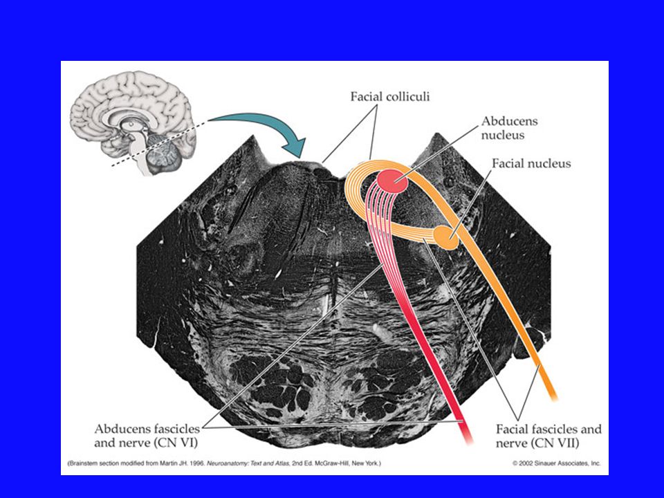
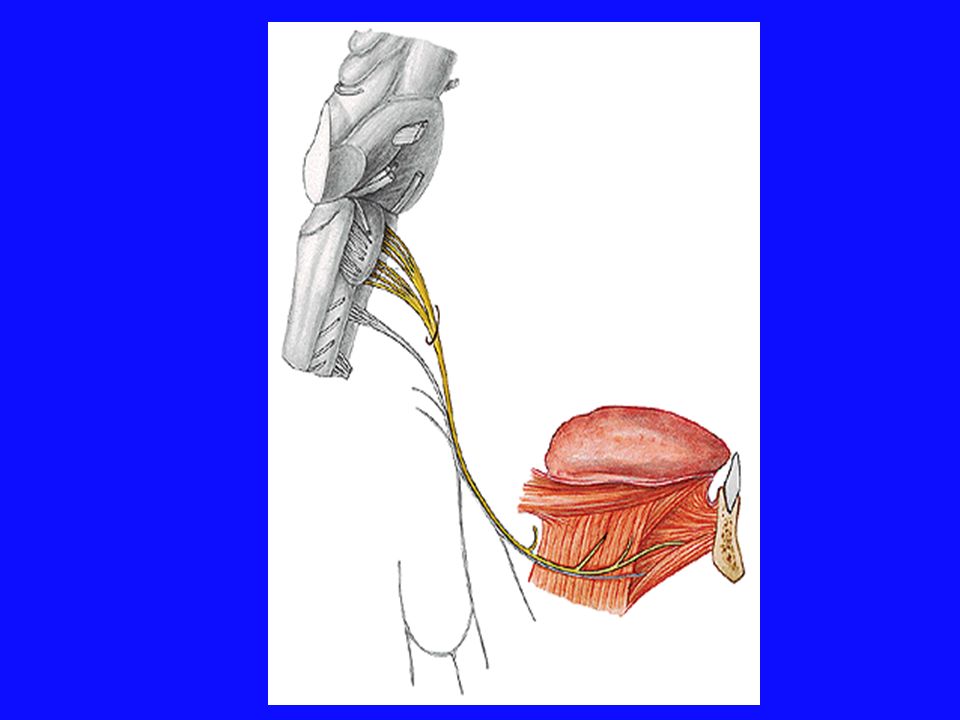
Nervus facialis N. intermedius Sulcus bulbopontinus
Motor ve duyu lifleri içeren miks bir sinir beyin sakını iki kök şeklinde terk eder Nervus facialis N. intermedius Sulcus bulbopontinus

3
ÖVE ÖVA GVE ÖVE Nucleus nervi facialis Motor Nuclei tractus solitarii
Nucleus salivatorius superior ÖVE ÖVA GVE ÖVE Nucleus nervi facialis

4
mezenkiminden gelişir
Mimik kaslarına somotomotor innervasyon M. digastricus’un arka karnı M. stylohyoideus M. stapedius ÖVE Sinirin büyük kısmını oluşturur Mimik kasları, 2. yutak kavsinin mezenkiminden gelişir

5
Visseromotor parasempatik
N. intermedius içinde nucleus salivatorius superior ‘dan başlayan ve Gl. lacrimalis Gl. submandibularis Gl. sublingualis Nasopharynx’teki bezler Sert damaktaki bezler giden GVE Preganglionik parasempatik lifler vardır Bazı bezlerin parasempatik inervasyonu sağlar

6
Meatus acusticus internus
Meatus acusticus internus ‘dan geçerek temporal kemiğin pars petrosa parçasındaki canalis facialis’ in içine girer geniculum’u oluşturur Ganglion geniculi GSA ve tat duyusunun birinci nöronu var Geniculum Ganglion Geniculi Canalis facialis Meatus acusticus internus Kafatasını terk eder

7
Canalis facialiste n. petrosus major n. stapedius
chorda tympani dallarını verir Canalis facialis’i ve cavum caranii ‘yi terk eder FORAMEN EUM

8
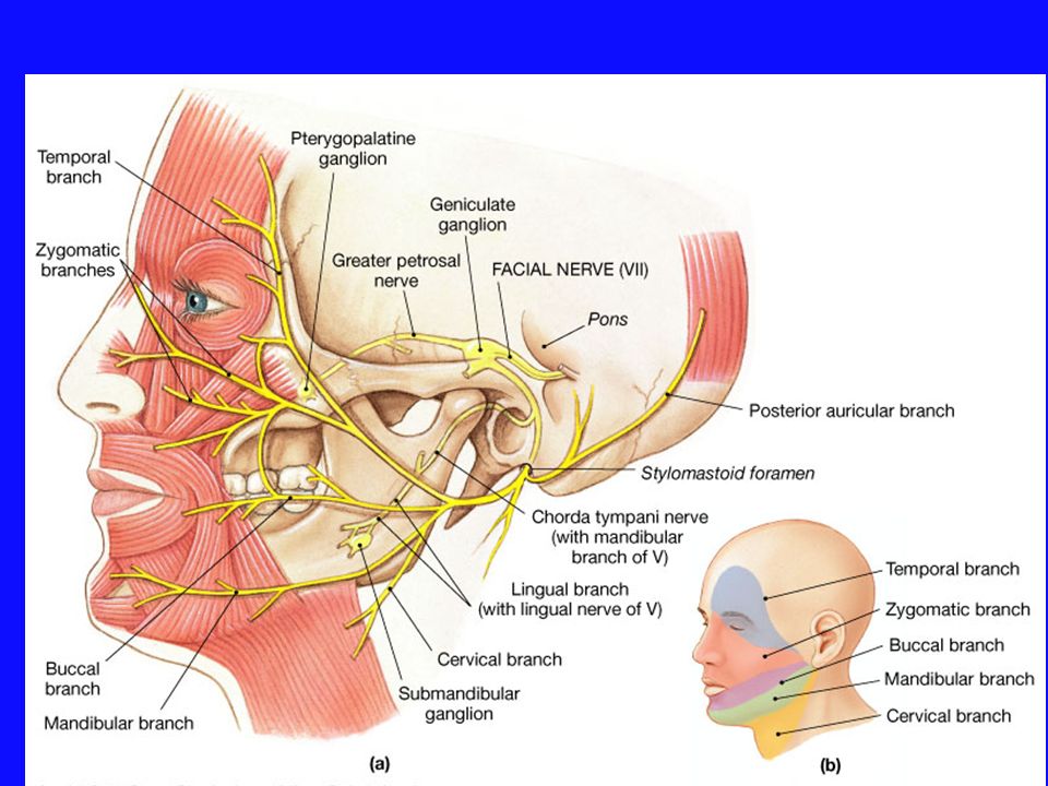
ÖVE Meatus acusticus internus Canalis facialis Rr. temporales
Somotomotor Brankiomotor Canalis facialis Rr. temporales Foramen stylomastoideum Rr. zygomatici Plexus intraparatideus Rr. buccales R. marginalis mandibulae Platysma M. digastricus’un arka karnı M. stylohyoideus M. stapedius R. colli (cervicalis)
")
9
Plexus intraparatideus

10
M. stylohyoideus M. digastricus’un arka karnı

11
Felcinde hiperacusia görülür
M. stapedius

14
N.petrosus major içerisinde damak mukozasından gelen tat duyusunu taşıyan lifler ile
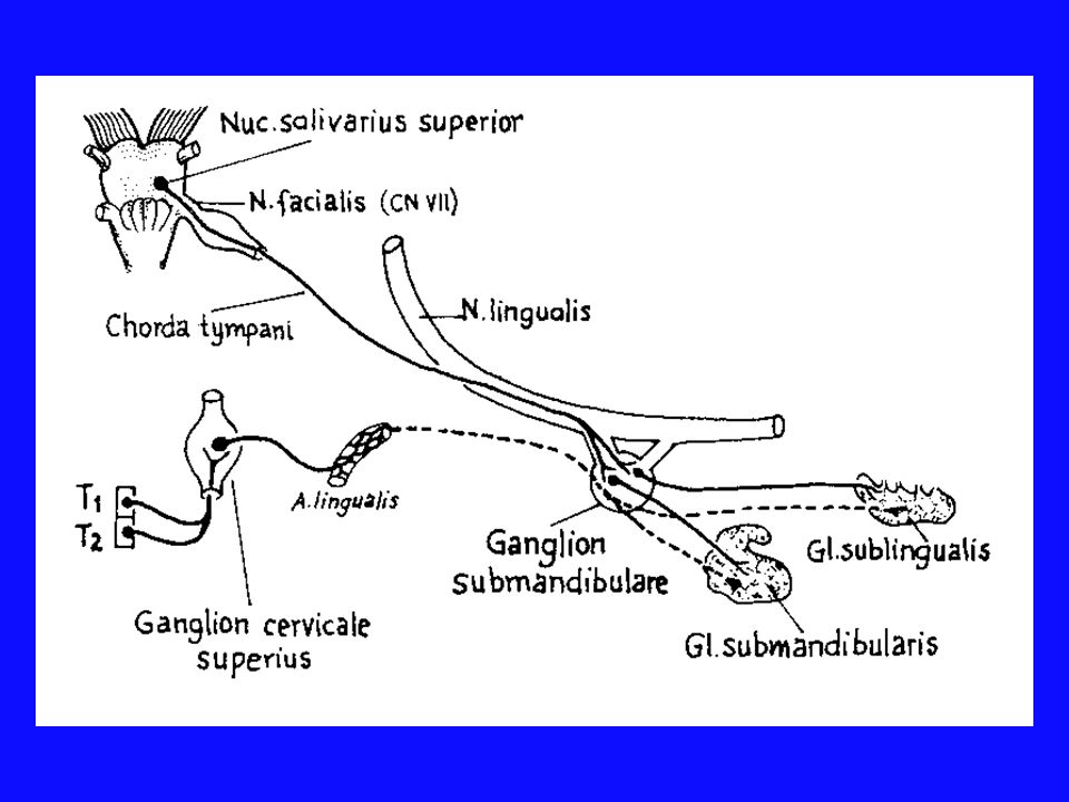
Nucleus salivatorius superior ganglion pterygopalatina başlayarak giden preganglionik parasempatik lifler bulunur N.petrosus major daha sonra Canalis pterygoidei içerisina girer ve n. canalis pterygoidei adını alır ve n. petrosus major ‘e sempatik lifler n.petrosus profundus ile plexus timpanicus’dan gelir ganglion pterygopalatina da sonlanır buradan başlayan postsinaptik parasempatik lifler n.maxillaris’ in r. zygomaticotemporalis dalı aracılığı ile n. lacrimalis’e katılır ve Gl. Lacrimalis Sekretomotor GVE inervasyonunu sağlar

15
Plexsus caroticus internus n.petrosus profundus
N.petrosus major daha sonra Canalis pterygoidei içerisina girer ve n. canalis pterygoidei adını alır ve Plexsus caroticus internus n.petrosus profundus n.canalis pterygoideii Vidius siniri

16
N. stapedius m. stapedius uyarır

17
Chorda tympani n.lingualis’e katılır Chorda tympani
Ganglion geniculi pseudounipolar nöronların periferik uzantıları Sentral uzantıları n.intermedius’a katılarak Nuclei solitarii gider N. facialis’ten canalis facialis içerisinde ayrılır içerisinde dilin 2/3 ön kısmından tat duyusu taşıyan lifler ve preganglionik parasempatik lifler bulunur Chorda tympani fossa infratemporalis’de n.lingualis’e katılır Nucleus salivatorius superior ‘dan başlar Ganglion submandibulare Gangliondan başlayan postganglionik parasempatik lifler Gl. submandibulare Gl. sublinguale

19
SOMATOSENSİTİF LİFLER
GSA Çeşitli bölgelerin somatik duyusunun alınması Kulak kepçesi Kulağın arkasında küçük bir alan Kulak zarının iç kısmı

20
Dilin 2/3 ön kısmından tat duyusu Yumuşak ve sert damak
ÖVA Chorda tympani Dilin 2/3 ön kısmından tat duyusu Yumuşak ve sert damak

21
Özel duyu tat ÖVA N. vagus N. glossopharyngeus N. facialis
Cranial Nerve IX - Glossopharyngeal Page 21 of 22 Overview of Special Sensory Component The special sensory component of CN IX provides taste sensation from the posterior one-third of the tongue., Peripheral course Special sensory fibers from the posterior one-third of the tongue travel via the pharyngeal branches of CN IX to the inferior glossopharyngeal ganglion where their cell bodies reside. Page 17 of 22 Peripheral course Sensory fibers from the skin of the external ear initially travel with the auricular branch of CN X, while those from the middle ear travel in the tympanic nerve as discussed above (CN IX visceral motor section). General sensory information from the upper pharynx and posterior one-third of the tongue travel via the pharyngeal branches of CN IX. These peripheral processes have cell their cell body in either the superior or inferior glossopharyngeal ganglion. Page 22 of 22 Central course - special sensory component The central processes of these neurons exit the inferior ganglion and pass through the jugular foramen to enter the brainstem at the level of the rostral medulla between the olive and inferior cerebellar peduncle. Upon entering the medulla these fibers ascend in the tractus solitarius and synapse in the caudal nucleus solitarius. Taste fibers from CN VII and X also ascend and synapse here. Figure 9-22a. Central course - special sensory component. Ascending secondary neurons originating in nucleus solitarius project bilaterally to the ventral posteromedial (VPM) nuclei of the thalamus via the central tegmental tract. Tertiary neurons from the thalamus project via the posterior limb of the internal capsule to the inferior one-third of the primary sensory cortex (the gustatory cortex of the parietal lobe). N. facialis (chorda tympani)
. General sensory information from the upper pharynx and posterior one-third of the tongue travel via the pharyngeal branches of CN IX. These peripheral processes have cell their cell body in either the superior or inferior glossopharyngeal ganglion. Page 22 of 22 Central course - special sensory component The central processes of these neurons exit the inferior ganglion and pass through the jugular foramen to enter the brainstem at the level of the rostral medulla between the olive and inferior cerebellar peduncle. Upon entering the medulla these fibers ascend in the tractus solitarius and synapse in the caudal nucleus solitarius. Taste fibers from CN VII and X also ascend and synapse here. Figure 9-22a. Central course - special sensory component. Ascending secondary neurons originating in nucleus solitarius project bilaterally to the ventral posteromedial (VPM) nuclei of the thalamus via the central tegmental tract. Tertiary neurons from the thalamus project via the posterior limb of the internal capsule to the inferior one-third of the primary sensory cortex (the gustatory cortex of the parietal lobe). N. facialis. (chorda tympani)")
22
Area gustatoria Alan 43

23
Cornea veya conjunctiva
Kornea refleksi Cornea veya conjunctiva’ya dokunulması göz kapaklarının kapanması ile sonuçlanan bir refleks harekete sebeb olur Cornea veya conjunctiva Alınan duyu impulsları Nucleus nervi facialis m.orbicularis oculi’yi İnerve eden nöronlarına iletir Buradaki inter nöronlar aldıkları impulsları MLF aracılığı ile Nucleus principalis nervi trigemini n.ophthalmicus

24
N.Facialis n.trigeminus’un n. ophthalmicus dalı Cornea refleksi
Afferent kısmı Cornea refleksi Efferent kısmı N.Facialis

25
SANTRAL TİP FASİAL PARALİZİ

26
Nucleus nervi facialis’e
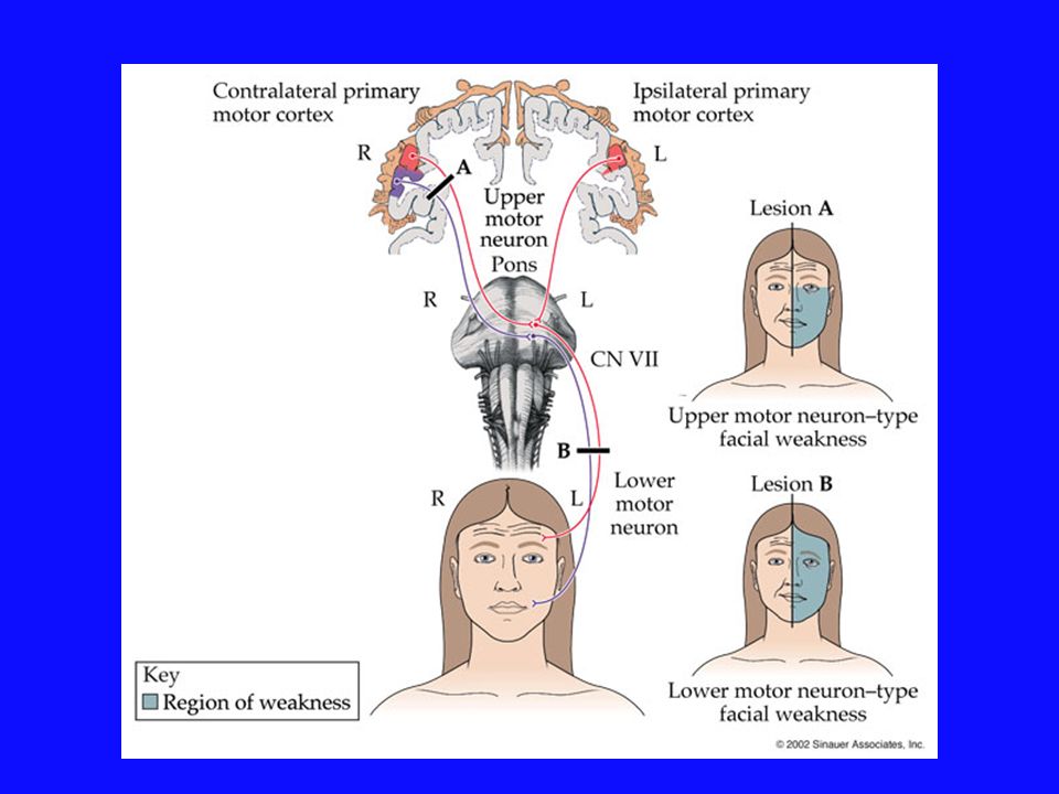
cortex’den fibrae corticonucleares ile gelen liflerin tek taraflı olarak zedelenmesi lezyonun karşı tarafında yüzün alt yarısında üst motor nöron paralizi ye santral facial paralizi sebeb olur n.facialis’in bu tür supranuclear lezyonlarında paralizinin sadece yüzün alt kısmında görülmesinin nedeni Nucleus nervi facialis’in yüzün alt yarısındaki kasları inerve eden kısmına yalnız kontralateral korteksten yüzün üst yarısındaki kasları inerve eden kısmına ise her iki korteksten lif gelmesidir

27
Lezyonun karşı tarafıda
Göz seviyesinin altında Ağız bölgesindeki kaslarda parezi veya paralizi görülür Ağız sağlam tarafa doğru kayar Göz ve alınla ilgili mimik kaslarında parezi veya paralizi görülmez Gözünü kapayabilir Alnını kırıştırabilir Kornea refleksi kaybolmaz

29
PERİFERİK TİP FASİAL PARALİZİ BELL PARALİZİ
N. facialis lezyonlarında ortaya çıkan tablo Beyin sakından çıktığı zaman zedelenirse ortaya çıkan semptomlar

30
Yüzün lezyon tarafındaki
yarımında Mimik kaslarda alt motor nöron tipi paralizi Hasta alnını kırıştıramaz Gözünü kapatamaz Dudaklarını büzemez Islık çalamaz Ağız bileşkesi aşağı sarkar Ağız sağlam tarafa kayar ** Kornea refleksi kaybolur ** Sublingual ve submandibular bezde fonksiyon bozukluğu **lakrimal bezde fonksiyon bozukluğu m.stapedius’un paralizine bağlı hyperacusis *** dil 2/3 ön kısmında tat duyusunda kayıp

31
**Yüzün lezyon tarafındaki
yarımında Mimik kaslarda alt motor nöron tipi paralizi Hasta alnını kırıştıramaz Gözünü kapatamaz Dudaklarını büzemez Islık çalamaz Ağız bileşkesi aşağı sarkar Ağız sağlam tarafa kayar ** Kornea refleksi kaybolur **M.stapedius’un paralizine bağlı hyperacusis Forame stylomastoideum’dan çıktığı yerde zedelenirse

32
Ganglion geniculi’in proksimalindeki n
Ganglion geniculi’in proksimalindeki n.facialis lezyonlarındaaksonların yanlış rejenerasyonu sonucu n.intermedius’taki preganglionik parasempatik lifler Chorda tympani yerine n.petrosus majör içerisine girer ve glandula lacrimalis’e gider Bu durumda kişi yemek yerken lezyon tarafındaki gözünden yaş akar Crocodile tears syndrome timsah göz yaşları sendromu

33
Stroke (right side) Bell palsy Santral tipte fasial sinir paralizisi
Periferik tipte fasial sinir paralizisi

34
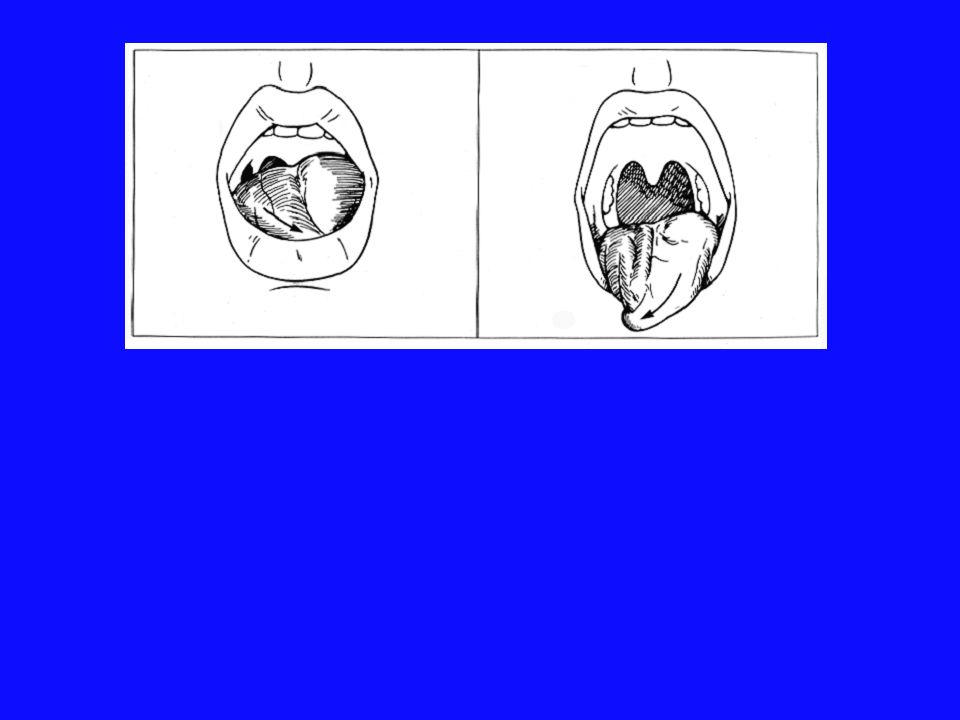
N. Facialis’in zedelenmesi sonucu Bell paralizisi (Bell’s palsy) denilen tablo ortaya çıkar.
Infranüklear fasial sinir paralizisi

35
ÖZET Miks bir sinirdir (motor, sensitif, parasempatik)
N. facialis Miks bir sinirdir (motor, sensitif, parasempatik) Mimik kaslarına somotomotor innervasyon sağlar Ganglion geniculi sensitif çekirdeğidir Chorda tympani adlı dalı dilin 2/3 ön kısmından tat duyusu alır ve Fissura petrotympanica’dan nevrax’a girer Parasempatik lifleri Gl. lacrimalis, Gl. sublingualis ve Gl. submandibularis’e innervasyon sağlar Parasempatik liflerin çekirdeği gang. pteryopalatinumdur Timsah ağlaması sendromu Bell paralizisi Santral tip fasiyal paralizi
 Mimik kaslarına somotomotor innervasyon sağlar. Ganglion geniculi sensitif çekirdeğidir. Chorda tympani adlı dalı dilin 2/3 ön kısmından tat duyusu alır ve Fissura petrotympanica’dan nevrax’a girer. Parasempatik lifleri Gl. lacrimalis, Gl. sublingualis ve Gl. submandibularis’e innervasyon sağlar. Parasempatik liflerin çekirdeği gang. pteryopalatinumdur. Timsah ağlaması sendromu. Bell paralizisi. Santral tip fasiyal paralizi.")
36
VIII. Nervus vestibulocochlearis

37
N. Vestibularis N. Cochlearis Sulcus bulbopontinus

38
N. Vestibularis SSA III.nöron CEREBELLUM I.nöron II.nöron Os temporale
Pars petrosa Meatus acusticus internus Crista ampullaris Central uzantısı Macula sacculi Ganglion vestibulare ( Scarpa ganglionu ) Bipolar nöronların N. Vestibularis I.nöron Macula utriculi Nucleus vestibularis superior Nucleus vestibularis lateralis Nucleus vestibularis medialis Nucleus vestibularis inferior II.nöron III.nöron CEREBELLUM
 Bipolar nöronların. N. Vestibularis. I.nöron. Macula utriculi. Nucleus vestibularis superior. Nucleus vestibularis lateralis. Nucleus vestibularis medialis. Nucleus vestibularis inferior. II.nöron. III.nöron. CEREBELLUM.")
39
N. Cochlearis İŞİTME YOLLARI LEMNİSCUS LATERALİS I.nöron II.nöron
Central uzantısı Ganglion spirale Bipolar nöronların Corti organı Organum spirale N. Cochlearis I.nöron Nucleus cochlearis ventralis Nucleus cochlearis dorsalis II.nöron İŞİTME YOLLARI LEMNİSCUS LATERALİS

41
IX. Nervus glossopharyngeus

42
ÖVE GVE GSA ÖVA GVA Branchiomotor kaslarına somotomotor innervasyon
M. stylopharyngeus Pharynx hareketleri Bazı bezlerin parasempatik innervasyonu Parotis bezi Larynx’teki düz kas ve bezler Pharynx’ teki düz kas ve bezler GVE Çeşitli bölgelerin somatik duyusunun alınması Dış kulak Kulak zarının iç yüzü Nasopharyx Dilin arka 1/3 bölümü GSA ÖVA Dilin 1/3 arka kısmından tat duyusu Sinus caroticus’taki baroreseptörler Glomus caroticum’daki kemoreseptörler GVA

44
The Glossopharyngeal Nerve
PLAY

45
Sulcus retroolivatis’ten beyni terk eder
Cranial Nerve IX - Glossopharyngeal Page 4 of 22 Rostral medulla Fibers leaving the nucleus ambiguus travel anteriorly and laterally to exit the medulla, along with the other components of CN IX, between the olive and the inferior cerebellar peduncle. Page 5 of 22 Intracranial Course - Branchial Motor Component Upon emerging from the lateral aspect of the medulla the branchial motor component joins the other components of CN IX to exit the skull via the jugular foramen. The glossopharyngeal fibers travel just anterior to the cranial nerves X and XI which also exit the skull via the jugular foramen. Page 6 of 22 Extra-cranial course and final innervation Upon exiting the skull the branchial motor fibers descend deep to the styloid process and wrap around the posterior border of the stylopharyngeus muscle before innervating it. N. glossopharyngeus Foramen jugulare’den nevrax’ı terk eder

46
Branchiomotor kaslarına somotomotor innervasyon M. stylopharyngeus
ÖVE Branchiomotor kaslarına somotomotor innervasyon M. stylopharyngeus Cranial Nerve IX - Glossopharyngeal Page 2 of 22 Overview of Branchial Motor Component The branchial motor component of CN IX provides voluntary control of the stylopharyngeus muscle which elevates the pharynx during swallowing and speech.

47
Somotomotor liflerin kaynağı formatio reticularis içerisinde bulunan Nucleus ambiguus’tur
Cranial Nerve IX - Glossopharyngeal Page 3 of 22 Origin and Central Course - Branchial Motor Component The branchial motor component originates from the nucleus ambiguus in the reticular formation of the medulla.

48
Branchiomotor kaslarına somotomotor innervasyon M. stylopharyngeus
Yutma ve konuşma sırasında Pharynx’i yukarı kaldırır M. stylopharyngeus

49
Larynx’teki düz kas ve bezler Pharynx’ teki düz kas ve bezler
GVE Parotis bezi Larynx’teki düz kas ve bezler Pharynx’ teki düz kas ve bezler parasempatik innervasyonu

50
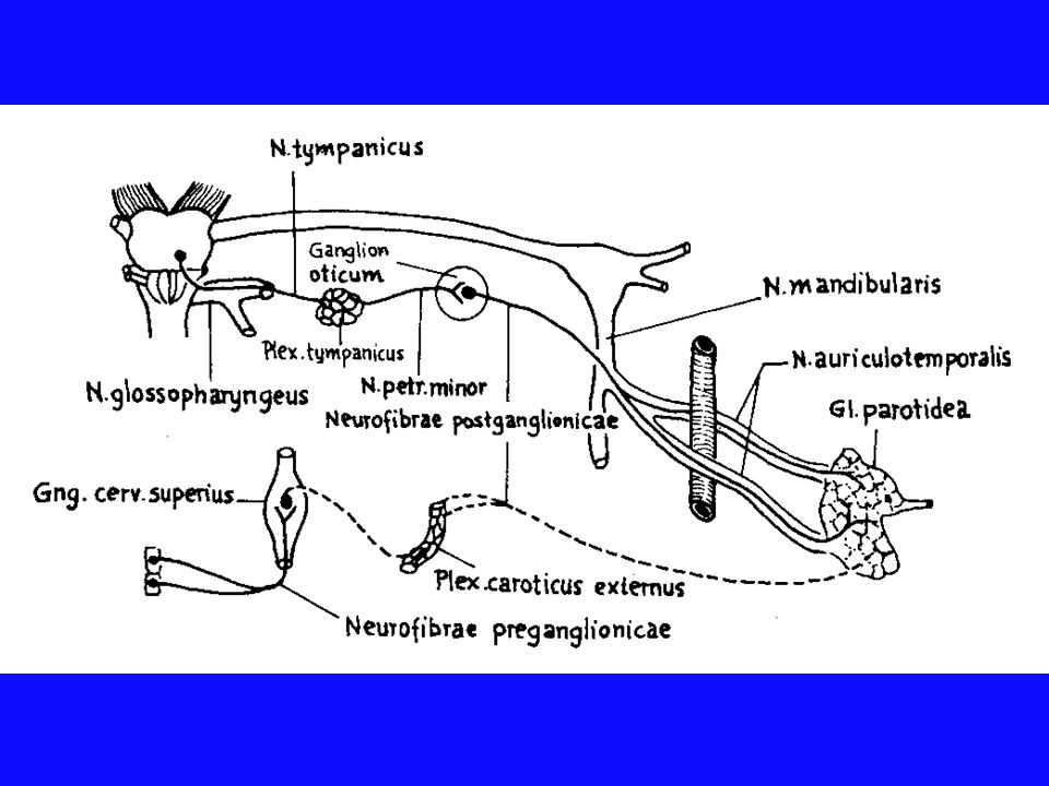
N. auriculotemporalis ganglion oticum N. Petrosus minor N . tympanicus
N. Glossopharyngeus içindeki GVE Ganglion inferius‘dan başlar Temporal kemikdeki canaliculus tympanicus Nucleus salivatorius inferior parasempatik liflerin kaynağı Plexus tympanicus promontorium N. Petrosus minor ganglion oticum Potsinaptik parasempatik lifler N. mandibularis N. auriculotemporalis GL. parotidea

52
GSA Çeşitli bölgelerin somatik duyusunun alınması Dış kulak
Kulak zarının iç yüzü Nasopharyx Dilin arka 1/3 bölümü Cranial Nerve IX - Glossopharyngeal Page 16 of 22 Overview of general sensory component This component of CN IX carries general sensory information (pain, temperature, and touch) from the skin of the external ear, internal surface of the tympanic membrane, the walls of the upper pharynx, and the posterior one-third of the tongue.
 from the skin of the external ear, internal surface of the tympanic membrane, the walls of the upper pharynx, and the posterior one-third of the tongue.")
53
Membrana tympani’nin sensitif innervasyonu
Dış yüzü N. vagus r. auricularis N. trigeminus n. auriculotemporalis İç yüz N. glossopharyngeus, N. facialis

54
Özel duyu (tat) Genel duyu ÖVA N. vagus N. glossopharyngeus
Dilin 1/3 arka kısmından tat duyusu Yumuşak ve sert damak Cranial Nerve IX - Glossopharyngeal Page 21 of 22 Overview of Special Sensory Component The special sensory component of CN IX provides taste sensation from the posterior one-third of the tongue., Peripheral course Special sensory fibers from the posterior one-third of the tongue travel via the pharyngeal branches of CN IX to the inferior glossopharyngeal ganglion where their cell bodies reside. Page 17 of 22 Peripheral course Sensory fibers from the skin of the external ear initially travel with the auricular branch of CN X, while those from the middle ear travel in the tympanic nerve as discussed above (CN IX visceral motor section). General sensory information from the upper pharynx and posterior one-third of the tongue travel via the pharyngeal branches of CN IX. These peripheral processes have cell their cell body in either the superior or inferior glossopharyngeal ganglion. Page 22 of 22 Central course - special sensory component The central processes of these neurons exit the inferior ganglion and pass through the jugular foramen to enter the brainstem at the level of the rostral medulla between the olive and inferior cerebellar peduncle. Upon entering the medulla these fibers ascend in the tractus solitarius and synapse in the caudal nucleus solitarius. Taste fibers from CN VII and X also ascend and synapse here. Figure 9-22a. Central course - special sensory component. Ascending secondary neurons originating in nucleus solitarius project bilaterally to the ventral posteromedial (VPM) nuclei of the thalamus via the central tegmental tract. Tertiary neurons from the thalamus project via the posterior limb of the internal capsule to the inferior one-third of the primary sensory cortex (the gustatory cortex of the parietal lobe). N. facialis (chorda tympani) N. trigeminus
. General sensory information from the upper pharynx and posterior one-third of the tongue travel via the pharyngeal branches of CN IX. These peripheral processes have cell their cell body in either the superior or inferior glossopharyngeal ganglion. Page 22 of 22 Central course - special sensory component The central processes of these neurons exit the inferior ganglion and pass through the jugular foramen to enter the brainstem at the level of the rostral medulla between the olive and inferior cerebellar peduncle. Upon entering the medulla these fibers ascend in the tractus solitarius and synapse in the caudal nucleus solitarius. Taste fibers from CN VII and X also ascend and synapse here. Figure 9-22a. Central course - special sensory component. Ascending secondary neurons originating in nucleus solitarius project bilaterally to the ventral posteromedial (VPM) nuclei of the thalamus via the central tegmental tract. Tertiary neurons from the thalamus project via the posterior limb of the internal capsule to the inferior one-third of the primary sensory cortex (the gustatory cortex of the parietal lobe). N. facialis. (chorda tympani) N. trigeminus.")
55
Glomus caroticum’daki kemoreseptörler
GVA Sinus caroticus’taki Baroreseptörler Glomus caroticum’daki kemoreseptörler Ramus sinus carotici (Hering siniri) Duyu taşır Cranial Nerve IX - Glossopharyngeal Page 13 of 22 Overview of visceral sensory component This component of CN IX innervates the baroreceptors of the carotid sinus and chemoreceptors of the carotid body.
 Duyu taşır. Cranial Nerve IX - Glossopharyngeal. Page 13 of 22 Overview of visceral sensory component This component of CN IX innervates the baroreceptors of the carotid sinus and chemoreceptors of the carotid body.")
56
Tr. solitarius sinus caroticus Foramen jugulare mmHg
Reticular formasyon, Hypothalamus’taki merkezler Foramen jugulare Kardiovasküler cevaplar (kan basıncı) Respiratuvar reflekler (CO2, O2) Gang. inf Cranial Nerve IX - Glossopharyngeal Page 14 of 22 Peripheral and intracranial course Sensory fibers arise from the carotid sinus and carotid body at the bifurcation of the common carotid artery, ascend in the sinus nerve, and join the other components of CN IX at the inferior hypoglossal ganglion. The cell bodies of these neurons reside in the inferior ganglion. The central processes of these neurons enter the skull via the jugular foramen. Page 15 of 22 Central course - visceral sensory component Once inside the skull, the visceral sensory fibers enter the lateral medulla between the olive and the inferior cerebellar peduncle and descend in the tractus solitarius to synapse in the caudal nucleus solitarius. From the nucleus solitarius, connections are made with several areas in the reticular formation and hypothalamus to mediate cardiovascular and respiratory reflex responses to changes in blood pressure, and serum concentrations of CO2 and O2. mmHg CO2 O2 sinus caroticus Glomus caroticum
 Respiratuvar reflekler (CO2, O2) Gang. inf. Cranial Nerve IX - Glossopharyngeal. Page 14 of 22 Peripheral and intracranial course Sensory fibers arise from the carotid sinus and carotid body at the bifurcation of the common carotid artery, ascend in the sinus nerve, and join the other components of CN IX at the inferior hypoglossal ganglion. The cell bodies of these neurons reside in the inferior ganglion. The central processes of these neurons enter the skull via the jugular foramen. Page 15 of 22 Central course - visceral sensory component Once inside the skull, the visceral sensory fibers enter the lateral medulla between the olive and the inferior cerebellar peduncle and descend in the tractus solitarius to synapse in the caudal nucleus solitarius. From the nucleus solitarius, connections are made with several areas in the reticular formation and hypothalamus to mediate cardiovascular and respiratory reflex responses to changes in blood pressure, and serum concentrations of CO2 and O2. mmHg. CO2. O2. sinus caroticus. Glomus caroticum.")
57
ÖZET N. glosopharyngeus
Miks bir sinirdir (motor, sensitif, parasempatik) Branchiomotor kasa “M. stylopharyngeus” giden lifler (ÖVE) Nucleus ambiguus’un üst kısmından başlar Foramen jugulareden nevrax’ı terk eder preganglionik parasempatik lifleri ganglion oticum’da sinaps yapar ve daha sonra gl. parotidea’ya innervasyon sağlar Dilin 1/3 arka kısmından tat ve genel duyu impulsları alır Baroreseptörlerden ve kemoreseptörlerden impulsları taşır
 Branchiomotor kasa M. stylopharyngeus giden lifler (ÖVE) Nucleus ambiguus’un üst kısmından başlar. Foramen jugulareden nevrax’ı terk eder. preganglionik parasempatik lifleri ganglion oticum’da sinaps yapar ve daha sonra gl. parotidea’ya innervasyon sağlar. Dilin 1/3 arka kısmından tat ve genel duyu impulsları alır. Baroreseptörlerden ve kemoreseptörlerden impulsları taşır.")
58
X. Nervus vagus

59
En uzun kraniyal sinir Üç nukleusu İki ganglionu var Ganglion superius jugulara Ganglion inferius nodosum

60
Ganglion superius Ganglion jugulara
GSA Ganglion superius Ganglion jugulara GSA liflerin I nöronları bulunur periferik uzantıları Kulağın arka kısmı Meatus acusticus externus Kulak zarının dış kısmı Pharynx Ganglion inferius Ganglion nodosum GVA liflerin I nöronları bulunur periferik uzantıları Larynx pharynx Toraks’taki organlar dan Abdomen’de flexura coli sinistra ‘ya kadar tüm sindirim kanalı organlarından visseral duyu taşır Arcus aorta’daki baroreseptörler Glomus aorticum’daki kemoreseptörler GVA

61
Ganglion inferius Ganglion nodosum
GVA liflerin I nöronları bulunur periferik uzantıları Epiglotis çevresinden tat duyusu ÖVA ÖVE Branchiomotor kaslarına somotomotor innervasyon Pharynx’in istemli kasları Larynx’in istemli kaslarının büyük çoğunluğu Dilin ekstrinsik kası M. palatoglossus GVE Bazı bezlerin parasempatik innervasyonu Larynx’teki düz kas ve bezler Pharynx’ teki düz kas ve bezler Toraks’taki organların innervasyonu Abdomen’deki organların innervasyonu Vagus" is from the Latin meaning wandering. This is a fitting name as the nerve wanders from the brainstem to the splenic flexure of the colon. The vagus nerve consists of five components with distinct functions:

62
R.Auricularis Arnold siniri R. pharyngeus N.laryngeus superior
R. Meningeus R.Auricularis Arnold siniri R. pharyngeus N.laryngeus superior Rr. Cardiaci cervicales superiores Rr. Cardiaci cervicales inferiores N.Laryngeus recurrens inferior PLAY

63
N. X Sulcus retroolivaris’ten ayrılır Nucleus ambiguus’un
orta kısmından kaynaklanır Cranial Nerve X - Vagus Page 3 of 24 Overview of Branchial Motor Component Origin and Central Course The branchial motor component originates from the nucleus ambiguus in the reticular formation of the medulla. Page 4 of 24 Rostral medulla Fibers leaving the nucleus ambiguus travel anteriorly and laterally to exit the medulla posterior to the olive as a series of rootlets. N. X

64
* Foramen jugulare içerisinde iken r. internus
Nervus vagus * N.Accessory r. internus Cranial Nerve X - Vagus Page 5 of 24 Intracranial Course Upon emerging from the lateral aspect of the medulla the branchial motor component travels with the fibers of the accessory nerve (CN XI) into the jugular foramen of the skull. The remaining components of the vagus nerve also enter the jugular foramen and give rise to two ganglia (the superior and inferior vagal ganglia) within the jugular foramen. The branchial motor fibers join with the rest of the vagus nerve just below the inferior vagal ganglion. Foramen jugulare içerisinde iken r. internus (kranial parçaya ait olan lifler) n. vagus’a katılır
 into the jugular foramen of the skull. The remaining components of the vagus nerve also enter the jugular foramen and give rise to two ganglia (the superior and inferior vagal ganglia) within the jugular foramen. The branchial motor fibers join with the rest of the vagus nerve just below the inferior vagal ganglion. Foramen jugulare içerisinde iken r. internus. (kranial parçaya ait olan lifler) n. vagus’a katılır.")
65
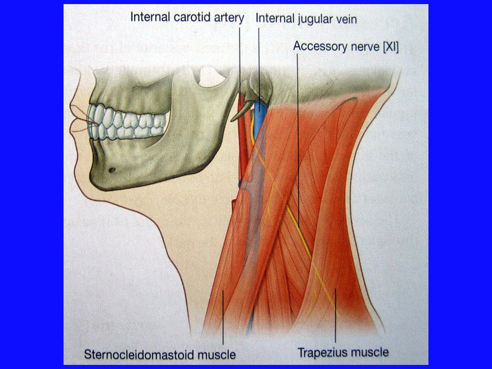
foramen jugulare N IX N X N XI V. jugularis interna
Cranial Nerve X - Vagus Page 6 of 24 Extra-Cranial Course and Final Innervation Upon exiting the skull the vagus nerve travels between the internal jugular vein and internal carotid artery within the carotid sheath. The branchial motor fibers leave the vagus nerve as three major branches: Pharyngeal branch Superior laryngeal nerve Recurrent laryngeal nerve foramen jugulare

66
Vagina carotica A. carotis interna v. jugularis interna N. V A G U S

67
XI. Nervus accessorius

68
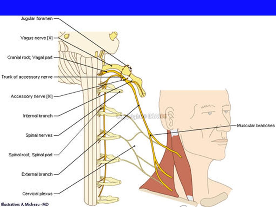
N. XI Sulcus retroolivaris’ten ayrılır
Cranial Nerve XI - Accessory Nerve Page 2 of 9 Origin and central course The fibers of the cranial root originate in the caudal nucleus ambiguus and travel anteriorly and laterally to exit the medulla between the olive and inferior cerebellar peduncle (just below the exiting fibers of CN X). N. XI
. N. XI.")
69
Motor bir sinirdir Kranial ve spinal olmak üzere iki kökü vardır

70
ÖVE Özel visseral efferent Special viscerel efferent
Branchiomotor kaslarına somotomotor inervasyon Larynx ve pharynx’in istemli kasları Kranial parça radix cranialis Branchiomotor kaslarına somotomotor inervasyon ÖVE M. sternocleidomastoideus ve m. trapezius’ un üst parçasını inerve eder spinal parça radix spinalis

71
* Accessory vagal nerve Kranial kökünü nucleus
Nervus vagus Kranial kökünü nucleus ambiquus içindeki nöronların aksonları yapar * Foramen jugulare ‘den Kafa dışına çıkınca kranial parçaya ait olan lifler r. internus n. vagus’a katılır Bu yüzden ramus cranialis’e pars vagalis adı da verilir ve n.vagus’un dalları içinde Larynx - pharynx ve yumuşak damak kaslarına gider Cranial Nerve XI - Accessory Nerve Page 3 of 9 Intracranial course Upon emerging from the medulla, the cranial root fibers momentarily join the spinal root fibers of CN XI and enter the jugular foramen. Within the foramen, the cranial root fibers split from the spinal root fibers and join the vagus nerve to exit the skull.

72
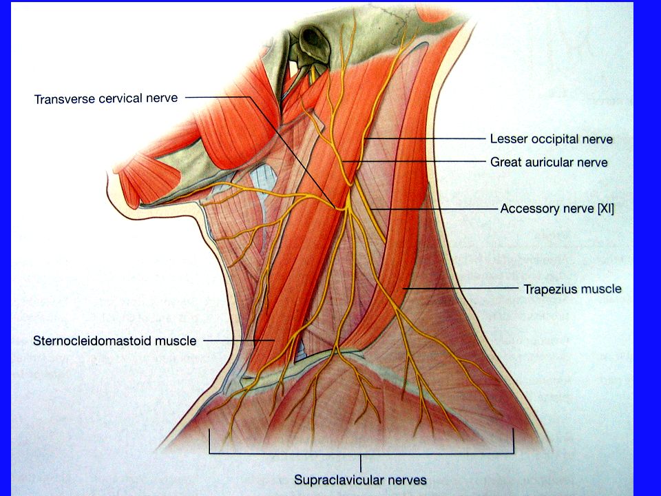
Spinal accessory nerve
Spinal kökü oluşturan lifler medulla spinalis’in ilk 5 segmentinde cornu anterior’da bulunan nöronların uzantıları meydana getirir Spinal kökün lifleri yukarı doğru yükselir foramen magnum’dan geçer Kranial kökün lifler ile birleşir Nervus vagus M. Trapezius M. sternocleidomastoideus

77
ÖZET N. accessorius Saf somotomotor bir sinirdir
Kranial bölümü Nucleus ambiguus’un alt kısmından başlar Spinal bölümü ise 1-5. medulla spinalis segmentlerinden kaynaklanır Kranial ve spinal bölüm birleştikten sonra foramen jugularede R. internus ve R. externus’a ayrılır R. internus, N. vagus’a katılır R. externus ise M. sternocleidomastoideus ve M. trapezius’a innervasyon sağlar Felcinde düşük omuz ve torticollis oluşur

78
XII. Nervus hypoglossus

80
Dil kaslarının motor siniri

81
Bulbus Beyin sakını sulcus anterolateralis’ ten sulcus preolivaris birkaç kök halinde terk eder

82
Nucleus motorius nervi hypoglossi Nervus hypoglossus

83
GSE M. digastricus venter posterior
Ocsipital kemikteki canalis nervi hypoglossi ‘den geçerek cavum cranii’ den çıkar a. Carotis externa ve dallarını dıştan çaprazlar N. hypoglossus A.carotis interna V. jugularis interna M. digastricus venter posterior Os hyoideum hizasında dil köküne girer

84
Ekstrinsik dil kaslarından m.hyoglossus m.styloglossus m.genioglossus
Bu kası uyaran nöronlar sadece karşı taraf korteksten Kortikonüklear lif alır Dil köküne girer İntrinsik dil kasları Ekstrinsik dil kaslarından m.hyoglossus m.styloglossus m.genioglossus m.palatoglossus hariç

85
Ganglion cervicale superius N. vagus ganglion inferius N
Ganglion cervicale superius N. vagus ganglion inferius N.lingualis bağlantıları vardır Nervus hypoglossus’dan ayrılan bir dal radix superior 1. ve 2. servikal spinal sinirlerden gelen bir dal radix inferior ile birleşir ve ansa cervicalis’i oluşturur Radix inferior Radix superior Musculi infrahyoidei

86
Tek taraflı XII. kraniyal sinir felcinde hasta dilini dışarı çıkardığında sağlam taraftaki m.geniglosus ‘un fonksiyonuna bağlı olarak dil lezyon tarafına doğru çıkar Dilin aynı yarısında atrofi görülür İki taraflı n. hypoglossus lezyonlarında dilin dışarı çıkarılmadığı, hatta ağız tabanında hiç hareket etmediği dikkati çeker

Benzer bir sunumlar


>")


>")


 Levent SARIKCIOĞLU.>")


>")