Sunuyu indir
Sunum yükleniyor. Lütfen bekleyiniz

1
Reseptörler ve Doz Yanıt İlişkisi
Prof. Dr. Ç. Hakan KARADAĞ

2
Reseptör Endojen maddelerin ya da ilaçların bağlanarak etkilerini oluşturdukları makromoleküllerdir. John Langley First postulated by John Langley (1878) Established after his experiments using nicotine and curare analogues on muscle contraction. Isolated muscle fibers: pilocarpine (contraction) and atropine (inhibition). Two compounds competing for a third, but unknown substrate. Furthered by Paul Ehrlich ( ) Demonstrated that stereoselectivity was imperative in drug-receptor signaling. In 1901, Langley challenged the dominant hypothesis that drugs act at nerve endings by demonstrating that nicotine acted at sympathetic ganglia even after the degeneration of the severed preganglionic nerve endings. That year, Langley also discovered for himself a tool in the form of renal extract (containing adrenaline) which produced sympathomimetic responses when applied to tissues exogenously. But it was not until 1905 that Langley published the results of the decisive experiments using systemic injections of curare and nicotine given to chicks. It was through these experiments that Langley concluded the existence of a receptive substance in striated muscle. Langley concluded that a protoplasmic "receptive substance" must exist which the two drugs compete for directly. He further added that the effect of combination of the receptive substance with competing drugs was determined by their comparative chemical affinities for the substance and relative dose. John N. Langley (1852–1925) P. Ehrlich (1854–1915) 2
 Established after his experiments using nicotine and curare analogues on muscle contraction. Isolated muscle fibers: pilocarpine (contraction) and atropine (inhibition). Two compounds competing for a third, but unknown substrate. Furthered by Paul Ehrlich ( ) Demonstrated that stereoselectivity was imperative in drug-receptor signaling. In 1901, Langley challenged the dominant hypothesis that drugs act at nerve endings by demonstrating that nicotine acted at sympathetic ganglia even after the degeneration of the severed preganglionic nerve endings. That year, Langley also discovered for himself a tool in the form of renal extract (containing adrenaline) which produced sympathomimetic responses when applied to tissues exogenously. But it was not until 1905 that Langley published the results of the decisive experiments using systemic injections of curare and nicotine given to chicks. It was through these experiments that Langley concluded the existence of a receptive substance in striated muscle. Langley concluded that a protoplasmic receptive substance must exist which the two drugs compete for directly. He further added that the effect of combination of the receptive substance with competing drugs was determined by their comparative chemical affinities for the substance and relative dose. John N. Langley (1852–1925) P. Ehrlich (1854–1915) 2.")
3
İntraselüler reseptörler
Steroid hormon reseptörleri Membranda yerleşmiş reseptörler İntraselüler kısmı enzim etkinliği gösteren reseptörler (tirozin kinaz) İntraselüler kısmı bir enzim ile kenetli reseptörler (tirozin kinaz) Yapılarında iyon kanalı bulunduran reseptörler (iyonotropik reseptör) G proteini ile kenetli reseptörler (metabotropik reseptör)
 İntraselüler kısmı bir enzim ile kenetli reseptörler (tirozin kinaz) Yapılarında iyon kanalı bulunduran reseptörler (iyonotropik reseptör) G proteini ile kenetli reseptörler (metabotropik reseptör)")
5
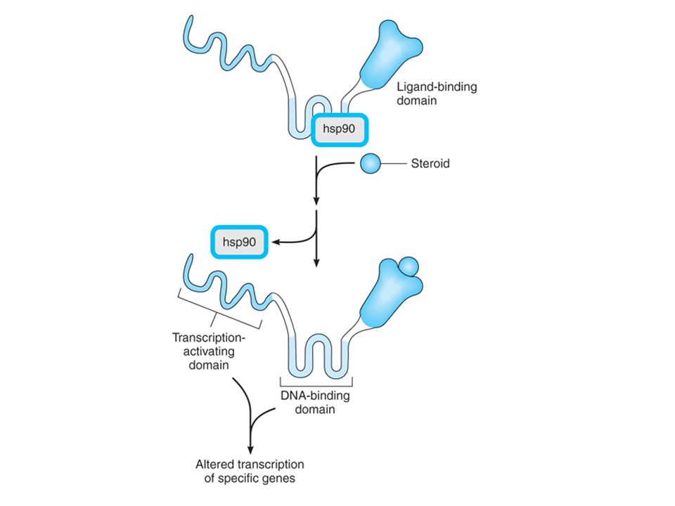
Steroid Reseptörünün Yapısı
İnhibitör protein kompleksi (hsp90) Hormon Transkripsiyonu aktive eden bölge DNA’ya bağlanan bölge Hormon bağlanma bölgesi
 Hormon. Transkripsiyonu aktive eden bölge. DNA’ya bağlanan bölge. Hormon bağlanma bölgesi.")
6
İntraselüler Yerleşimli Reseptör

7
Hormon Yanıt Elemanları (HRE)
Promoter Hormon Yanıt Elemanı (HRE) Transkripsiyon Başlangıç Kompleksi Hormon ya da Efektör HRE Glukokortikoidler GRE Projestinler PRE Mineralokortikoidler MRE Androjenler ARE Estrojenler ERE Tiroid hormon TRE Retinoik asit RARE Vitamin D VDRE
 Transkripsiyon Başlangıç Kompleksi. Hormon ya da Efektör HRE. Glukokortikoidler GRE. Projestinler PRE. Mineralokortikoidler MRE. Androjenler ARE. Estrojenler ERE. Tiroid hormon TRE. Retinoik asit RARE. Vitamin D VDRE.")
8
İntraselüler reseptörler
Steroid hormon reseptörleri Membranda yerleşmiş reseptörler İntraselüler kısmı enzim etkinliği gösteren reseptörler (tirozin kinaz) İntraselüler kısmı bir enzim ile kenetli reseptörler (tirozin kinaz) Yapılarında iyon kanalı bulunduran reseptörler (iyonotropik reseptör) G proteini ile kenetli reseptörler (metabotropik reseptör)
 İntraselüler kısmı bir enzim ile kenetli reseptörler (tirozin kinaz) Yapılarında iyon kanalı bulunduran reseptörler (iyonotropik reseptör) G proteini ile kenetli reseptörler (metabotropik reseptör)")
9
Epidermal Büyüme Faktörü (EGF)
Mechanism of activation of the epidermal growth factor (EGF) receptor, a representative receptor tyrosine kinase. The receptor polypeptide has extracellular and cytoplasmic domains, depicted above and below the plasma membrane. Upon binding of EGF (circle), the receptor converts from its inactive monomeric state (left) to an active dimeric state (right), in which two receptor polypeptides bind noncovalently. The cytoplasmic domains become phosphorylated (P) on specific tyrosine residues (Y) and their enzymatic activities are activated, catalyzing phosphorylation of substrate proteins (S). Ligand-Regulated Transmembrane Enzymes Including Receptor Tyrosine Kinases This class of receptor molecules mediates the first steps in signaling by insulin, epidermal growth factor (EGF), platelet-derived growth factor (PDGF), atrial natriuretic peptide (ANP), transforming growth factor-b (TGF-b), and many other trophic hormones. These receptors are polypeptides consisting of an extracellular hormone-binding domain and a cytoplasmic enzyme domain, which may be a protein tyrosine kinase, a serine kinase, or a guanylyl cyclase (Figure 2-7). In all these receptors, the two domains are connected by a hydrophobic segment of the polypeptide that crosses the lipid bilayer of the plasma membrane. The receptor tyrosine kinase signaling pathway begins with binding of ligand, typically a polypeptide hormone or growth factor, to the receptor's extracellular domain. The resulting change in receptor conformation causes receptor molecules to bind to one another, which in turn brings together the tyrosine kinase domains, which become enzymatically active, and phosphorylate one another as well as additional downstream signaling proteins. Activated receptors catalyze phosphorylation of tyrosine residues on different target signaling proteins, thereby allowing a single type of activated receptor to modulate a number of biochemical processes. Insulin, for example, uses a single class of receptors to trigger increased uptake of glucose and amino acids, and to regulate metabolism of glycogen and triglycerides in the cell. Similarly, each of the growth factors initiates in its specific target cells a complex program of cellular events ranging from altered membrane transport of ions and metabolites to changes in the expression of many genes. Inhibitors of receptor tyrosine kinases are finding increased use in neoplastic disorders where excessive growth factor signaling is often involved. Some of these inhibitors are monoclonal antibodies (eg, trastuzumab, cetuximab), which bind to the extracellular domain of a particular receptor and interfere with binding of growth factor. Other inhibitors are membrane-permeant "small molecule" chemicals (eg, gefitinib, erlotinib), which inhibit the receptor's kinase activity in the cytoplasm. The intensity and duration of action of EGF, PDGF, and other agents that act via receptor tyrosine kinases are limited by a process called receptor down-regulation. Ligand binding often induces accelerated endocytosis of receptors from the cell surface, followed by the degradation of those receptors (and their bound ligands). When this process occurs at a rate faster than de novo synthesis of receptors, the total number of cell-surface receptors is reduced (down-regulated) and the cell's responsiveness to ligand is correspondingly diminished. A well-understood example is the EGF receptor tyrosine kinase, which undergoes rapid endocytosis and is trafficked to lysosomes after EGF binding; genetic mutations that interfere with this process cause excessive growth factor-induced cell proliferation and are associated with an increased susceptibility to certain types of cancer. Endocytosis of other receptor tyrosine kinases, most notably receptors for nerve growth factor, serves a very different function. Internalized nerve growth factor receptors are not rapidly degraded and are translocated in endocytic vesicles from the distal axon, where receptors are activated by nerve growth factor released from the innervated tissue, to the cell body. In the cell body the growth factor signal is transduced to transcription factors regulating the expression of genes controlling cell survival. This process effectively transports a critical survival signal from its site of release to its site of signaling effect, and does so over a remarkably long distance¾more than 1 meter in certain sensory neurons. A number of regulators of growth and differentiation, including TGF-b, act on another class of transmembrane receptor enzymes that phosphorylate serine and threonine residues. ANP, an important regulator of blood volume and vascular tone, acts on a transmembrane receptor whose intracellular domain, a guanylyl cyclase, generates cGMP (see below). Receptors in both groups, like the receptor tyrosine kinases, are active in their dimeric forms. İnsülin Epidermal Büyüme Faktörü (EGF) Trombosit Kaynaklı Büyüme Faktörü (PDGF) Sinir Büyüme Faktörü (NGF) İnsülin-benzeri Büyüme Faktörü-1 (IGF-1) 9
 receptor, a representative receptor tyrosine kinase. The receptor polypeptide has extracellular and cytoplasmic domains, depicted above and below the plasma membrane. Upon binding of EGF (circle), the receptor converts from its inactive monomeric state (left) to an active dimeric state (right), in which two receptor polypeptides bind noncovalently. The cytoplasmic domains become phosphorylated (P) on specific tyrosine residues (Y) and their enzymatic activities are activated, catalyzing phosphorylation of substrate proteins (S). Ligand-Regulated Transmembrane Enzymes Including Receptor Tyrosine Kinases This class of receptor molecules mediates the first steps in signaling by insulin, epidermal growth factor (EGF), platelet-derived growth factor (PDGF), atrial natriuretic peptide (ANP), transforming growth factor-b (TGF-b), and many other trophic hormones. These receptors are polypeptides consisting of an extracellular hormone-binding domain and a cytoplasmic enzyme domain, which may be a protein tyrosine kinase, a serine kinase, or a guanylyl cyclase (Figure 2-7). In all these receptors, the two domains are connected by a hydrophobic segment of the polypeptide that crosses the lipid bilayer of the plasma membrane. The receptor tyrosine kinase signaling pathway begins with binding of ligand, typically a polypeptide hormone or growth factor, to the receptor s extracellular domain. The resulting change in receptor conformation causes receptor molecules to bind to one another, which in turn brings together the tyrosine kinase domains, which become enzymatically active, and phosphorylate one another as well as additional downstream signaling proteins. Activated receptors catalyze phosphorylation of tyrosine residues on different target signaling proteins, thereby allowing a single type of activated receptor to modulate a number of biochemical processes. Insulin, for example, uses a single class of receptors to trigger increased uptake of glucose and amino acids, and to regulate metabolism of glycogen and triglycerides in the cell. Similarly, each of the growth factors initiates in its specific target cells a complex program of cellular events ranging from altered membrane transport of ions and metabolites to changes in the expression of many genes. Inhibitors of receptor tyrosine kinases are finding increased use in neoplastic disorders where excessive growth factor signaling is often involved. Some of these inhibitors are monoclonal antibodies (eg, trastuzumab, cetuximab), which bind to the extracellular domain of a particular receptor and interfere with binding of growth factor. Other inhibitors are membrane-permeant small molecule chemicals (eg, gefitinib, erlotinib), which inhibit the receptor s kinase activity in the cytoplasm. The intensity and duration of action of EGF, PDGF, and other agents that act via receptor tyrosine kinases are limited by a process called receptor down-regulation. Ligand binding often induces accelerated endocytosis of receptors from the cell surface, followed by the degradation of those receptors (and their bound ligands). When this process occurs at a rate faster than de novo synthesis of receptors, the total number of cell-surface receptors is reduced (down-regulated) and the cell s responsiveness to ligand is correspondingly diminished. A well-understood example is the EGF receptor tyrosine kinase, which undergoes rapid endocytosis and is trafficked to lysosomes after EGF binding; genetic mutations that interfere with this process cause excessive growth factor-induced cell proliferation and are associated with an increased susceptibility to certain types of cancer. Endocytosis of other receptor tyrosine kinases, most notably receptors for nerve growth factor, serves a very different function. Internalized nerve growth factor receptors are not rapidly degraded and are translocated in endocytic vesicles from the distal axon, where receptors are activated by nerve growth factor released from the innervated tissue, to the cell body. In the cell body the growth factor signal is transduced to transcription factors regulating the expression of genes controlling cell survival. This process effectively transports a critical survival signal from its site of release to its site of signaling effect, and does so over a remarkably long distance¾more than 1 meter in certain sensory neurons. A number of regulators of growth and differentiation, including TGF-b, act on another class of transmembrane receptor enzymes that phosphorylate serine and threonine residues. ANP, an important regulator of blood volume and vascular tone, acts on a transmembrane receptor whose intracellular domain, a guanylyl cyclase, generates cGMP (see below). Receptors in both groups, like the receptor tyrosine kinases, are active in their dimeric forms. İnsülin. Epidermal Büyüme Faktörü (EGF) Trombosit Kaynaklı Büyüme Faktörü (PDGF) Sinir Büyüme Faktörü (NGF) İnsülin-benzeri Büyüme Faktörü-1 (IGF-1) 9.")
10
Receptor protein-tyrosine kinases transmit signals across the plasma membrane, from the cell exterior to the cytoplasm. The interaction of the external domain of a receptor tyrosine kinase with the ligand, often a growth factor, up-regulates the enzymatic activity of the intracellular catalytic domain, which causes tyrosine phosphorylation of cytoplasmic signaling molecules. 10

11
İntraselüler Kısmı Enzim Etkinliği Gösteren Reseptör

12
İntraselüler reseptörler
Steroid hormon reseptörleri Membranda yerleşmiş reseptörler İntraselüler kısmı enzim etkinliği gösteren reseptörler (tirozin kinaz) İntraselüler kısmı bir enzim ile kenetli reseptörler (tirozin kinaz) Yapılarında iyon kanalı bulunduran reseptörler (iyonotropik reseptör) G proteini ile kenetli reseptörler (metabotropik reseptör)
 İntraselüler kısmı bir enzim ile kenetli reseptörler (tirozin kinaz) Yapılarında iyon kanalı bulunduran reseptörler (iyonotropik reseptör) G proteini ile kenetli reseptörler (metabotropik reseptör)")
13
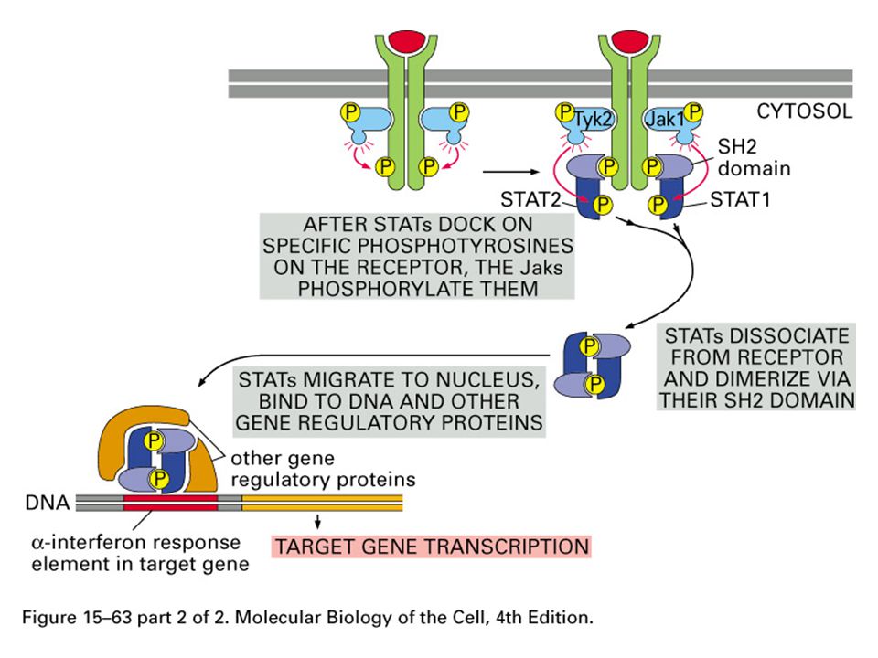
Koloni stimüle edici faktörler
Cytokine receptors, like receptor tyrosine kinases, have extracellular and intracellular domains and form dimers. However, after activation by an appropriate ligand, separate mobile protein tyrosine kinase molecules (JAK) are activated, resulting in phosphorylation of signal transducers and activation of transcription (STAT) molecules. STAT dimers then travel to the nucleus, where they regulate transcription. 0 Cytokine Receptors Cytokine receptors respond to a heterogeneous group of peptide ligands that includes growth hormone, erythropoietin, several kinds of interferon, and other regulators of growth and differentiation. These receptors use a mechanism (Figure 2-8) closely resembling that of receptor tyrosine kinases, except that in this case, the protein tyrosine kinase activity is not intrinsic to the receptor molecule. Instead, a separate protein tyrosine kinase, from the Janus-kinase (JAK) family, binds noncovalently to the receptor. As in the case of the EGF-receptor, cytokine receptors dimerize after they bind the activating ligand, allowing the bound JAKs to become activated and to phosphorylate tyrosine residues on the receptor. Tyrosine phosphates on the receptor then set in motion a complex signaling dance by binding another set of proteins, called STATs (signal transducers and activators of transcription). The bound STATs are themselves phosphorylated by the JAKs, two STAT molecules dimerize (attaching to one another's tyrosine phosphates), and finally the STAT/STAT dimer dissociates from the receptor and travels to the nucleus, where it regulates transcription of specific genes. Büyüme hormonu Eritropoietin Sitokinler Koloni stimüle edici faktörler JAK : Janus kinase STAT : Signal Transducers and Activators of Transcription 13
 are activated, resulting in phosphorylation of signal transducers and activation of transcription (STAT) molecules. STAT dimers then travel to the nucleus, where they regulate transcription. 0. Cytokine Receptors Cytokine receptors respond to a heterogeneous group of peptide ligands that includes growth hormone, erythropoietin, several kinds of interferon, and other regulators of growth and differentiation. These receptors use a mechanism (Figure 2-8) closely resembling that of receptor tyrosine kinases, except that in this case, the protein tyrosine kinase activity is not intrinsic to the receptor molecule. Instead, a separate protein tyrosine kinase, from the Janus-kinase (JAK) family, binds noncovalently to the receptor. As in the case of the EGF-receptor, cytokine receptors dimerize after they bind the activating ligand, allowing the bound JAKs to become activated and to phosphorylate tyrosine residues on the receptor. Tyrosine phosphates on the receptor then set in motion a complex signaling dance by binding another set of proteins, called STATs (signal transducers and activators of transcription). The bound STATs are themselves phosphorylated by the JAKs, two STAT molecules dimerize (attaching to one another s tyrosine phosphates), and finally the STAT/STAT dimer dissociates from the receptor and travels to the nucleus, where it regulates transcription of specific genes. Büyüme hormonu. Eritropoietin. Sitokinler. Koloni stimüle edici faktörler. JAK : Janus kinase STAT : Signal Transducers and Activators of Transcription. 13.")
15
İntraselüler reseptörler
Steroid hormon reseptörleri Membranda yerleşmiş reseptörler İntraselüler kısmı enzim etkinliği gösteren reseptörler (tirozin kinaz) İntraselüler kısmı bir enzim ile kenetli reseptörler (tirozin kinaz) Yapılarında iyon kanalı bulunduran reseptörler (iyonotropik reseptör) G proteini ile kenetli reseptörler (metabotropik reseptör)
 İntraselüler kısmı bir enzim ile kenetli reseptörler (tirozin kinaz) Yapılarında iyon kanalı bulunduran reseptörler (iyonotropik reseptör) G proteini ile kenetli reseptörler (metabotropik reseptör)")
16
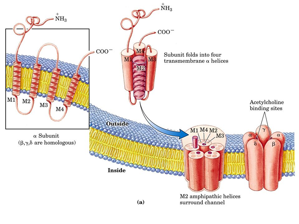
Asetilkolin nikotinik
GABAA Glutamat (NMDA, AMPA) Glisin Eksitatör (EPSP) Na+ Ca2+ İnhibitör (IPSP) Cl-
 Glisin. Eksitatör (EPSP) Na+ Ca2+ İnhibitör (IPSP) Cl-")
18
İyon Kanalı ile Kenetli Reseptörler

19
İntraselüler reseptörler
Steroid hormon reseptörleri Membranda yerleşmiş reseptörler İntraselüler kısmı enzim etkinliği gösteren reseptörler (tirozin kinaz) İntraselüler kısmı bir enzim ile kenetli reseptörler (tirozin kinaz) Yapılarında iyon kanalı bulunduran reseptörler (iyonotropik reseptör) G proteini ile kenetli reseptörler (metabotropik reseptör)
 İntraselüler kısmı bir enzim ile kenetli reseptörler (tirozin kinaz) Yapılarında iyon kanalı bulunduran reseptörler (iyonotropik reseptör) G proteini ile kenetli reseptörler (metabotropik reseptör)")
24
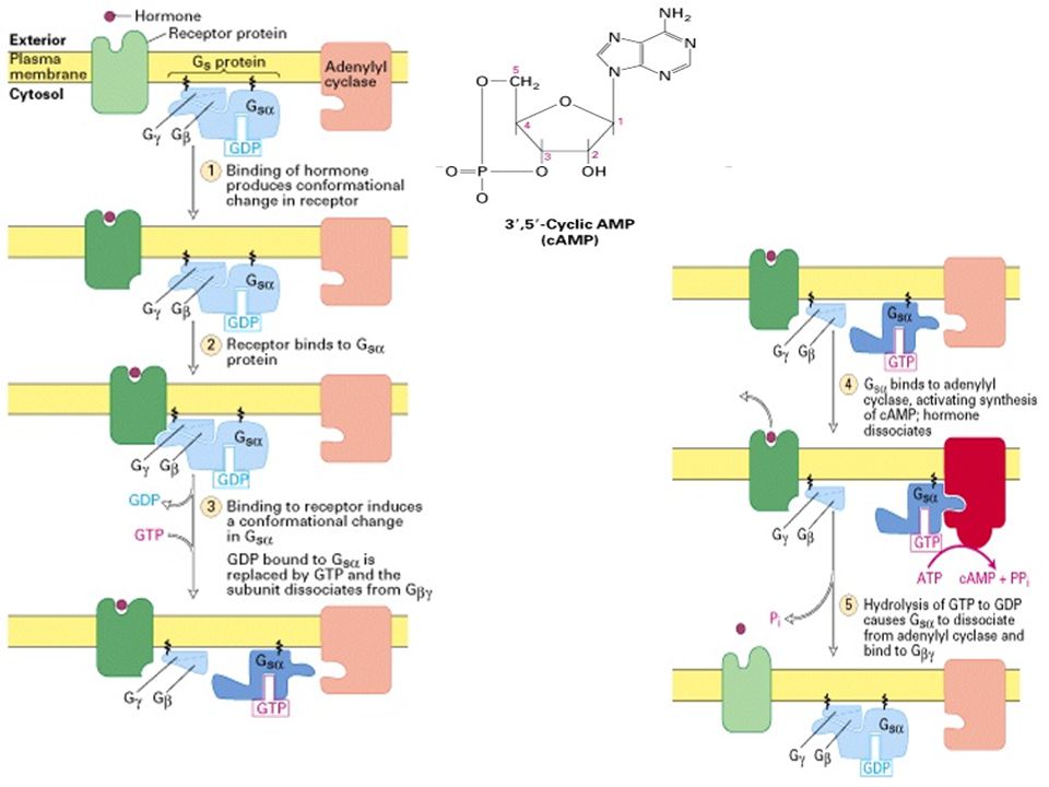
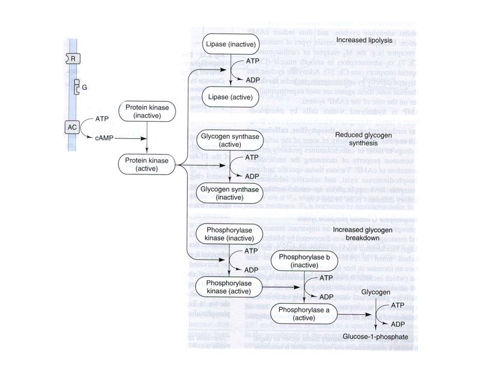
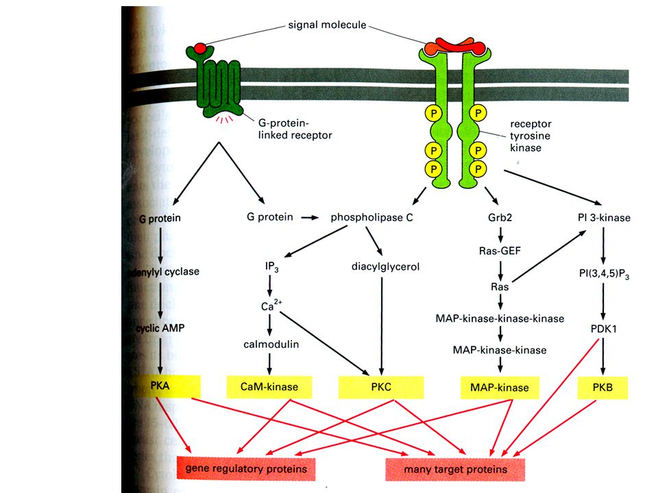
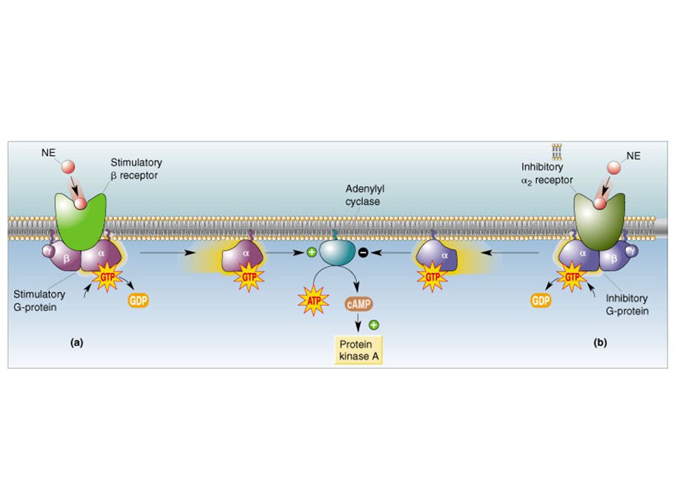
Adenilil siklaz aktivasyonu ve hücre içinde sAMP artışı

27
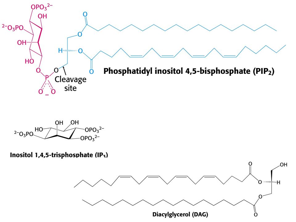
PIP2. : Fosfatidil İnositol Bifosfat DAG. : Diasilgliserol IP3
PIP2 : Fosfatidil İnositol Bifosfat DAG : Diasilgliserol IP3 : İnositol Trifosfat

31
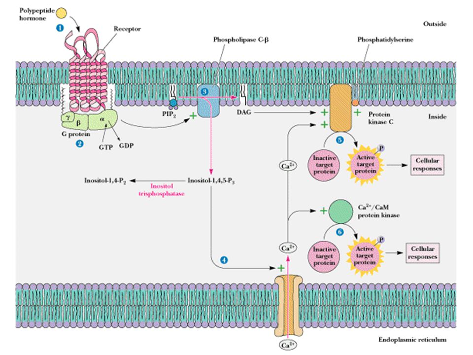
Fosfolipaz C aktivasyonu ve hücre içinde inositol trifosfat (IP3) ve diasilgliserol (DAG) artışı
 ve diasilgliserol (DAG) artışı")
33
Düzenleyici Protein Tipleri
Gs Adenilil siklaz ↑ sAMP ↑ beta adrenerjik reseptörler glukagon, histamin, serotonin Gi1, Gi2, Gi3 Adenilil siklaz ↓ sAMP ↓ K+ kanallarının açılması a2-adrenerjik reseptörler, asetilkolin (muskarinik), opioidler, serotonin G0 ? ? SSS (spesifik olarak belirlenmemiş) Golf Adenilil siklaz ↑ sAMP ↑ Kokular Gq Fosfolipaz C ↑ IP3 ↑ DAG↑ asetilkolin (muskarinik), serotonin (5-HT1C), a1-adrenerjik reseptörler Gt1, Gt2 sGMP fosfodiesteraz ↑ sGMP ↓ fotonlar (retinal hücrelerde rodopsin ve opsinler)
, opioidler, serotonin. G0 SSS (spesifik olarak belirlenmemiş) Golf Adenilil siklaz ↑ sAMP ↑ Kokular. Gq Fosfolipaz C ↑ IP3 ↑ DAG↑ asetilkolin (muskarinik), serotonin (5-HT1C), a1-adrenerjik reseptörler. Gt1, Gt2 sGMP fosfodiesteraz ↑ sGMP ↓ fotonlar (retinal hücrelerde rodopsin ve opsinler)")
35
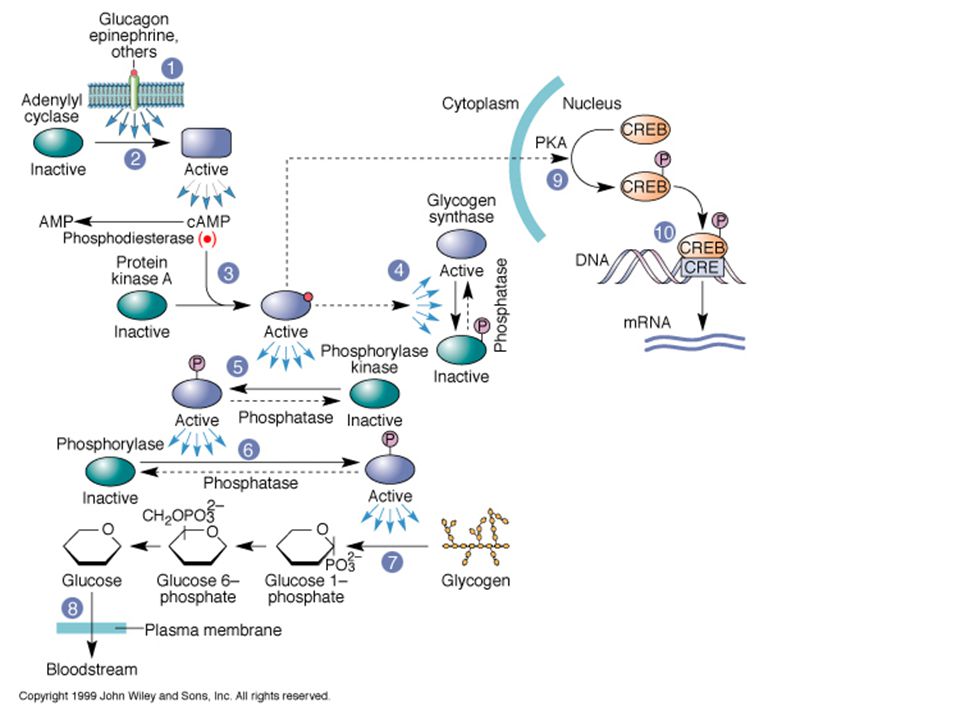
Sinyal Transdükleme Yanıtın Güçlendirilmesini (Amplifikasyon) Sağlar
 Sağlar")
36
İntraselüler reseptörler
Steroid hormon reseptörleri Membranda yerleşmiş reseptörler İntraselüler kısmı enzim etkinliği gösteren reseptörler (tirozin kinaz) İntraselüler kısmı bir enzim ile kenetli reseptörler (tirozin kinaz) Yapılarında iyon kanalı bulunduran reseptörler (iyonotropik reseptör) G proteini ile kenetli reseptörler (metabotropik reseptör)
 İntraselüler kısmı bir enzim ile kenetli reseptörler (tirozin kinaz) Yapılarında iyon kanalı bulunduran reseptörler (iyonotropik reseptör) G proteini ile kenetli reseptörler (metabotropik reseptör)")
37
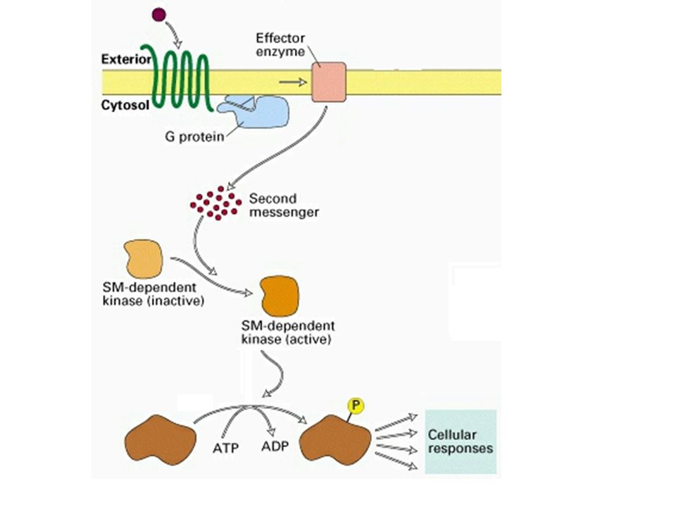
Sinyal İletiminin Önemi
The Nobel Prize in Physiology or Medicine 1994 "for their discovery of G-proteins and the role of these proteins in signal transduction in cells" Alfred G. Gilman Martin Rodbell USA University of Texas, Southwestern Medical Center Dallas, TX, USA National Institute of Environmental Health Sciences Research Triangle Park, NC, USA 1941 -

38
Sinyal İletiminin Önemi
The Nobel Prize in Physiology or Medicine 2000 "signal transduction in the nervous system" Arvid Carlsson Paul Greengard Eric R Kandel Sweden USA Göteborg University Göteborg, Sweden Rockefeller University New York, NY, USA Columbia University New York, NY, USA 1923 - 1925 - 1929 -

39
İlaç – Reseptör Etkileşmesinin Kinetiği ve Doz – Yanıt İlişkisi

40
Agonist Antagonist Ligand
Reseptöre bağlanarak bir etki oluşturabilen maddelere agonist denir. Antagonist Reseptöre bağlanan, doğrudan bir etki oluşturmayan, ancak agonistin bağlanmasını engelleyen maddelere antagonist denir. Ligand Reseptöre bağlanabilen maddelere (agonist ya da antagonist olabilir) ligand denir.
 ligand denir.")
41
1E-08 1E-6 0.0001 0.0005 0.001

42
Disosiasyon sabitesi Hill-Langmuir Eşitliği

43
Reseptörlerin yarısı işgal edildiğinde denklem ne duruma gelir?
Hill-Langmuir Eşitliği Archibald Vivian Hill Maksimum etkinin yarısına erişildiğinde reseptörlerin yarısı işgal edilmiş durumdadır. Bu durumu sağlayan ilaç konsantrasyonu değeri, ilacın disosiasyon sabitesine eşittir. (???)
")
44
Emaks (efikasite) EC50 : 1 x 10⁻⁶ M pD2 : 6 EC50
 EC50 : 1 x 10⁻⁶ M pD2 : 6 EC50")
45
ED50 EC50 TD50 LD50

46
Gravimetrik güç A>B>C
Efikasite A=B=C

47
Gravimetrik güç D>A>B>C>E A B C Efikasite A=B=C>D=E
A, B ve C tam agonist D ve E parsiyel agonist

48
Parsiyel Agonist

49
Parsiyel Agonist Efikasitesi standart agonistten (tam agonistten) daha düşük olan agonistlerdir. Ortamda tam agonist yok iken agonist gibi davranır. Ortamda tam agonist varken antagonist gibi davranır.

50
Agonist Antagonist

51
Yedek Reseptörler Parsiyel Agonist

53
Tolerans Sürekli kullanılan ilaçlara karşı yanıtın giderek azalmasıdır. Biyokimyasal veya farmakokinetik tolerans Otoindüksiyon Farmakodinamik veya hücresel tolerans Down-regulation Up-regulation

54
Taşifilaksi Toleransın çok çabuk gelişen şeklidir. Akut toleransdır.
Taşifilaksiye yol açan maddeler genellikle vücutta endojen bir maddeyi salıvererek etki oluştururlar. Efedrin, tiramin, amfetamin vb. Desensitizasyon da (duyarsızlama) bir taşifilaksi şeklidir.
 bir taşifilaksi şeklidir.")
55
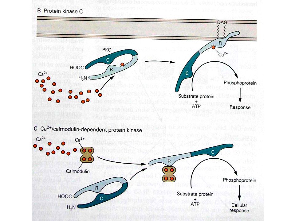
Desensitizasyon Agonist ortamda bulunduğu halde yanıtın azalması durumudur. Olası Mekanizmalar Reseptör konformasyonunun bozulması Reseptör molekülünün kovalent modifikasyonu Sekestrasyon Reseptör-G protein kenetlenmesinin bozulması GTP-G proteini kenetlenmesinin bozulması İkinci ulak düzeyinde adaptif değişiklikler.

57
Antagonizma Kimyasal antagonizma kelat (şelat) demir – desferrioksamin
Fizyolojik antagonizma glukagon – insulin Farmakolojik antagonizma Kompetitif antagonizma Kompetitif-olmayan antagonizma 57

58
Kompetitif antagonizma
+10-6M Ant +10-5M Ant

60
Kompetitif-olmayan antagonizma
+10-6M Ant +10-5M Ant

62
log (r – 1) = log [B] – log KB
10 mM 100 mM 1000 mM 2.698 -3 499 500 1000 1.690 -4 49 50 100 0.699 -5 5 6 10 - 1 Log [r –1] Log [anta] r – 1 dose ratio (r) [agonist] (mM) [anta] 3 Schild Grafiği 2 Schild denklemi log (r – 1) = log [B] – log KB log (r-1) 1 log KB (pA2) Heinz Otto Schild 1906 – 1984 -6.0 -5.5 -5.0 -4.5 -4.0 -3.5 -3.0 -2.5 -2.0 log [antagonist] KB değerinin negatif logaritması (-log KB) pA2 değerini verir.
![log (r – 1) = log [B] – log KB](http://slideplayer.biz.tr/slide/3076322/11/images/62/log+%28r+%E2%80%93+1%29+%3D+log+%5BB%5D+%E2%80%93+log+KB.jpg "10 mM. 100 mM mM Log [r –1] Log [anta] r – 1. dose ratio (r) [agonist] (mM) [anta] 3. Schild Grafiği. 2. Schild denklemi. log (r – 1) = log [B] – log KB. log (r-1) 1. log KB (pA2) Heinz Otto Schild – log [antagonist] KB değerinin negatif logaritması (-log KB) pA2 değerini verir.")
63
LD50 ED50 100 Etki (%) 50% doz (veya log doz) Terapötik İndeks (Tİ)
Terapötik Etki Letal Doz Etki (%) 50% Median Etkin Doz - ED50 Medyan Letal Doz - LD50 doz (veya log doz)
 50% Median Etkin Doz - ED50. Medyan Letal Doz - LD50. doz (veya log doz)")
64
Etkinin maksimuma erişme süresi
Minimum toksik konsantrasyon [C]p plazma doruk konsantrasyonu GÜVENLİK ARALIĞI (terapötik pencere) Etkinin başlama süresi Etki süresi Minimum etkin konsantrasyon Etkinin maksimuma erişme süresi Zaman
 Etkinin başlama süresi. Etki süresi. Minimum etkin konsantrasyon. Etkinin maksimuma erişme süresi. Zaman.")
Benzer bir sunumlar