Sunuyu indir
Sunum yükleniyor. Lütfen bekleyiniz

1
Hİsteroskopİk Tubal Kateterİzasyon
Dr. Ümit GÖKTOLGA 2nci Güncel Üreme Endokrinolojisi ve YÜT Kongresi Nisan 2008 İZMİR

2
Hİsteroskopİk Tubal Kateterİzasyon Nedİr ?
Asıl olarak proksimal tubal obstrüksyonların tedavisinde Minimal invaziv yöntem 1847 de Smith ve 1856 da Gardner ilk olarak kör tubal kanulasyon uygulamasını gerçekleştirmiştir.

3
Uygulama alanlarI Proksimal Tubal oklüzyon İnseminasyon
Gamet ve embriyo transferi için intratubal ortamı değerlendirmek için kullanılabilir. Tubal sterilizasyon Servikal stenoz IVF öncesi Hydrosalpenks tedavisi Tubal gebelik tedavisi

4
Uterus anatomİsİ

5
Tuba Uterİna İntramural veya interstisyel İsthmus Ampulla İnfundibulum
Fimbria

6
İntramural Kısım 1,5-2,5 cm uzunluğundadır.
Tüp bu seviyede dardır. (0,8-1,2 mm) Epiteliel hasar olmaksızın 1,0-1,2 mm lik kanülün geçişine için verir. Seyri değişken olabilir. Genellikle utero tubal junctiona doğru düz veya hafif kurve lü olabilir. The normal intramural oviduct ranges from 1.5 – 2.5 cms in length and takes a straight to slightly curved course to the utero tubal junction. It is 0.8 – 1.2 mm in diameter and can accommodate a cannula of mm in diameter without epithelial damage. Each ostium is situated at the apex of the uterotubal gutter and can be seen hysteroscopically at the bottom of a saucer-shaped depression as a sharp membranous ring which measures 0.8 – 1.2 mm in diameter. Therefore, if one passes a flexible, smooth-walled, J-shaped directional cannula through the internal os and directs it towards the left or right, it will follow the utero-tubal gutter towards the respective tubal ostia. A description and illustration of the passage of a wire probe through the intramural segment of the human oviduct using a transvaginal approach was first published by Gardener in Since then bioengineers have custom designed and miniaturized guidewires, cannulas & flexible endoscopes. Less flexible cannulas and endoscopes will tend to get stuck in the intramural bulge as it narrows about 1-1.5cms. beyond the ostia. Hence the importance of over the wire or coaxial cannulation systems to keep the lumen coaxially aligned with the tubal lumen, for advancement. Smaller cannulas with diameter of 0.6mm may negotiate most intramural lumens without coaxial assistance. Greater attention to coaxial alignment is necessary during hysteroscopic cannulation techniques as the distension medium converts the uterine cavity into a three-dimensional structure. The isthmic portion of oviduct commonly takes a degree bend as it extends beyond the uterotubal junction and is a potential site for tubal perforation.
 Epiteliel hasar olmaksızın 1,0-1,2 mm lik kanülün geçişine için verir. Seyri değişken olabilir. Genellikle utero tubal junctiona doğru düz veya hafif kurve lü olabilir. The normal intramural oviduct ranges from 1.5 – 2.5 cms in length and takes a straight to slightly curved course to the utero tubal junction. It is 0.8 – 1.2 mm in diameter and can accommodate a cannula of mm in diameter without epithelial damage. Each ostium is situated at the apex of the uterotubal gutter and can be seen hysteroscopically at the bottom of a saucer-shaped depression as a sharp membranous ring which measures 0.8 – 1.2 mm in diameter. Therefore, if one passes a flexible, smooth-walled, J-shaped directional cannula through the internal os and directs it towards the left or right, it will follow the utero-tubal gutter towards the respective tubal ostia. A description and illustration of the passage of a wire probe through the intramural segment of the human oviduct using a transvaginal approach was first published by Gardener in Since then bioengineers have custom designed and miniaturized guidewires, cannulas & flexible endoscopes. Less flexible cannulas and endoscopes will tend to get stuck in the intramural bulge as it narrows about 1-1.5cms. beyond the ostia. Hence the importance of over the wire or coaxial cannulation systems to keep the lumen coaxially aligned with the tubal lumen, for advancement. Smaller cannulas with diameter of 0.6mm may negotiate most intramural lumens without coaxial assistance. Greater attention to coaxial alignment is necessary during hysteroscopic cannulation techniques as the distension medium converts the uterine cavity into a three-dimensional structure. The isthmic portion of oviduct commonly takes a degree bend as it extends beyond the uterotubal junction and is a potential site for tubal perforation.")
7
İsthmus Ampulla İnfindubulum 3-4 cm uzunluğundadır. Çapı 0,5-1mm dir.

8
İntramural Fallop Tüpü
Zengin inervasyon ve vaskülarizasyona sahiptir. Hormonal Medikal Nörolojik uyarılara duyarlıdır.

9
İntramural Fallop Tüpü
Siliasyonu Epitel yüksekliğini Mitotik aktiviteyi Fallop tüpünün kontraksiyonunu Östrojen Progesteron

10
İntramural Tubal Patolojİ
En önemlisi Tubal Oklüzyonlardır. Salpenjit Fibrozis Endometriozis Tubal polipler Debris veya proteinöz materyale sekonder psödooklüzyon

11
Hİsteroskopİk Tubal KanÜlasyon
Rijit ve flexible operatif histeroskop 7 mm dış çaplı 7F operating kanal Aşırı anteflex veya retroflex durumda 4,9 mm dış çaplı En uygun sıvı % 5 dextroz ve ringer laktat Kanulasyon seti: Rijit histeroskop; Novy kanulasyon seti Flexible histeroskopta; Katayoma katateri

12
Teknİk GAA da Monitörizasyon için laparoskopi gereklidir.
Önce laparoskop yerleştirilerek pelvis değerlendirilir. Histeroskopiye geçilir. Serviks dilate edilir. Işık kaynağı Genişletici medium

13
Teknİk Tubal ostiuma katater yerleştirilir.
Utero-tubal yollar izlenir. İç katater tubal açıklığa yakın kısma kadar ilerletilir. Öne-arkaya hareket ettirilerek atravmatik olarak tüplere girilebilir. Direnç mevcutsa işlemden vazgeçilir. Tubal fibrozis ?

14
Teknİk Kornual segment klavuz tel ile kanüle edilince, 3fr katateri klavuz tel üzerinde kaydırılır ve klavuz çıkarılır. Bu katatere indigo-karmen enjekte edilir. Laparoskop ile geçiş izlenir. Geçiş doğrulanınca aynı işlem kontralateral tüpte gerçekleştirilir.

15
İşlemden sonra 3 gün süre ile
Uterin kavitede anatomik varyasyon veya tubal açıklıkta angulasyon mevcut ise, Flexible histeroskop tercih edilmelidir. İşlemden 30 dk önce 1 gr cefazolin Na İşlemden sonra 3 gün süre ile Sefaleksin tb 4x1

16
Proksİmal Tubal Oklüzyon
İnfertilite nedenlerinin % 25 ini tubal faktör oluşturmaktadır. Tubal blokaj Tubal blokaj olmaksızın tubal skar oluşumu ve diğer tubal hasarlar % si ise proksimal fallop tüpü obstrüksyonudur. Sulak et al 1987

17
Proksİmal Tubal Oklüzyon
Nedenleri Uterin fibroidler Peritubal adezyonlar İnfeksiyonlar İntratubal hastalıklar (endometriozis, salpenjitis vs) Sonuçları İntraluminal debris İntraluminal adezyon Stenozis Fibrozis
 Sonuçları. İntraluminal debris. İntraluminal adezyon. Stenozis. Fibrozis.")
18
Proksİmal Tubal Oklüzyon
Başarı oranı yaklaşık % 75 tir. (11/15) Histeroskopik olarak tedavi edilen hastalarda gebelik oranı % 13,3 Okluzyon ortadan kaldırılan hastalarda 3 aylık izlem sonrası HSG ile komplet patens oranı % 78 dir. Spiewankiewicz B et al. Clin Exp Obstet Gynecol 1995
 Histeroskopik olarak tedavi edilen hastalarda gebelik oranı % 13,3. Okluzyon ortadan kaldırılan hastalarda 3 aylık izlem sonrası HSG ile komplet patens oranı % 78 dir. Spiewankiewicz B et al. Clin Exp Obstet Gynecol")
19
Proksİmal Tubal oklüzyon
Histeroskopik tubal kanulasyon (Grup 1) ile rezeksiyon anastomoz u (Grup 2) karşılaştıran retrospektif çalışma Her grupta eşit sayıda toplam 74 hasta; İntrauterin gebelik oranları benzer % 57 vs % 50 Ektopik gebelik oranı daha düşük % 0 vs % 29 Das K et al. Fertil Steril 1995
 ile rezeksiyon anastomoz u (Grup 2) karşılaştıran retrospektif çalışma. Her grupta eşit sayıda toplam 74 hasta; İntrauterin gebelik oranları benzer. % 57 vs % 50. Ektopik gebelik oranı daha düşük. % 0 vs % 29. Das K et al. Fertil Steril")
20
Proksİmal Tubal Oklüzyon
Retrospektif çalışma 29 proksimal tubal oklüzyonlu hasta 13 hastada başarı ile kanülasyon sağlandı. (% 45) 3 hasta takipten çıkmış 10 hastadan 2 term de doğum 1 ektopik gebelik 7 olgu IVF e yönlendirilmiş. Maikis r et al. Gynaecological Endoscopy, 2000 Uzun dönem sonuçları ??????? Gynaecological Endoscopy Volume 9 Issue 6 Page , December 2000 To cite this article: Roseann Maikis, Ted L. Anderson, James F. Daniell (2000) Hysteroscopic tubal cannulation: long-term results Gynaecological Endoscopy 9 (6) , 397–400 doi: /j x Prev Article Next Article Abstract Hysteroscopic tubal cannulation: long-term results Roseann Maikis,1 1 Department of Obstetrics and Gynecology, University of Virginia, Charlottesville, Virginia, USA Ted L. Anderson2 2 Department of Obstetrics and Gynecology, Centennial Medical Center, Nashville, Tennessee, USA and James F. Daniell2 2 Department of Obstetrics and Gynecology, Centennial Medical Center, Nashville, Tennessee, USA 1 Department of Obstetrics and Gynecology, University of Virginia, Charlottesville, Virginia, USA 2 Department of Obstetrics and Gynecology, Centennial Medical Center, Nashville, Tennessee, USA CorrespondenceJ.F. Daniell, 2222 State Street, Suite A, Nashville, TN 37203, USA. Objective To examine the success rate and long-term results of hysteroscopic cannulation under laparoscopic control for proximal fallopian tube obstruction. Design Retrospective outcome analysis. Setting Referral endoscopic private practice for infertility. Subjects 29 patients referred with proximal tubal cornual obstruction, confirmed by hysterosalpingography or laparoscopy, with other tubal factors confounding infertility. All patients underwent hysteroscopic proximal guide wire tubal cannulation under laparoscopic guidance. Main outcome measures Pregnancy outcomes from 2 to 5 years following the procedure. Results Of a total of 29 attempts at tubal cannulation, 16 were unsuccessful at the time of surgery (55%), while 13 patients had successful proximal tubal cannulation, and patency confirmed via intraoperative chromotubation. Of these 13, three were lost to long-term follow up. Among the 10 remaining patients, there were three pregnancies: two term deliveries and one ectopic pregnancy. The remaining seven of the 10 patients with successful cannulations did not achieve pregnancy (70%) and elected for in vitro fertilization or adoption. Conclusion After over a decade of reports of hysteroscopic tubal cannulation, this procedure has been used as the first-line, and often only, therapy for proximal tubal obstruction. The results of this series indicate that pregnancy rates are not as favourable as reported by others. These long-term poor results should be considered when counselling patients with cornual occlusion about this surgery or other assisted reproductive options.
 3 hasta takipten çıkmış. 10 hastadan. 2 term de doğum. 1 ektopik gebelik. 7 olgu IVF e yönlendirilmiş. Maikis r et al. Gynaecological Endoscopy, Uzun dönem sonuçları Gynaecological Endoscopy. Volume 9 Issue 6 Page , December To cite this article: Roseann Maikis, Ted L. Anderson, James F. Daniell (2000) Hysteroscopic tubal cannulation: long-term results Gynaecological Endoscopy 9 (6) , 397–400 doi: /j x. Prev Article Next Article. Abstract. Hysteroscopic tubal cannulation: long-term results. Roseann Maikis,1 1 Department of Obstetrics and Gynecology, University of Virginia, Charlottesville, Virginia, USA. Ted L. Anderson2 2 Department of Obstetrics and Gynecology, Centennial Medical Center, Nashville, Tennessee, USA and. James F. Daniell2 2 Department of Obstetrics and Gynecology, Centennial Medical Center, Nashville, Tennessee, USA. 1 Department of Obstetrics and Gynecology, University of Virginia, Charlottesville, Virginia, USA 2 Department of Obstetrics and Gynecology, Centennial Medical Center, Nashville, Tennessee, USA. CorrespondenceJ.F. Daniell, 2222 State Street, Suite A, Nashville, TN 37203, USA. Objective To examine the success rate and long-term results of hysteroscopic cannulation under laparoscopic control for proximal fallopian tube obstruction. Design Retrospective outcome analysis. Setting Referral endoscopic private practice for infertility. Subjects 29 patients referred with proximal tubal cornual obstruction, confirmed by hysterosalpingography or laparoscopy, with other tubal factors confounding infertility. All patients underwent hysteroscopic proximal guide wire tubal cannulation under laparoscopic guidance. Main outcome measures Pregnancy outcomes from 2 to 5 years following the procedure. Results Of a total of 29 attempts at tubal cannulation, 16 were unsuccessful at the time of surgery (55%), while 13 patients had successful proximal tubal cannulation, and patency confirmed via intraoperative chromotubation. Of these 13, three were lost to long-term follow up. Among the 10 remaining patients, there were three pregnancies: two term deliveries and one ectopic pregnancy. The remaining seven of the 10 patients with successful cannulations did not achieve pregnancy (70%) and elected for in vitro fertilization or adoption. Conclusion After over a decade of reports of hysteroscopic tubal cannulation, this procedure has been used as the first-line, and often only, therapy for proximal tubal obstruction. The results of this series indicate that pregnancy rates are not as favourable as reported by others. These long-term poor results should be considered when counselling patients with cornual occlusion about this surgery or other assisted reproductive options.")
21
Tubal patens 1 yıl içerisinde kontrol edilmelidir.
Tubal reoklüzyon oranı % dur. Histeroskopik tubal kanulasyonun başarı oranı % dur. İntrauterin gebelik oranı % dir. Ektopik gebelik oranı % 8 dir.

22
Alternatİf Yöntemler Fluoroskopik tubal kanulasyon
Radyologlar tercih eder. HSG ile aynı teknik. HSG ile uterin kavitenin taslağı ve kornuların lokalizasyonu ortaya çıkartılır. Koaksial katater sistemi doğrudan intramural segmente doğru yayılır.

23
3F katateri 5F kataterin içine yerleştirilir ve çapı 0,5 mm den daha küçük olan klavuz tel geçirilir. Bu tel obstrükte alana kadar iletilir ve klavuz telin üzerinde 3F katateri kaydırılır. Klavuz tel çıkartılır ve kontrast madde direkt tubal lümene enjekte edilir.

24
Tubal kanulasyon başarılınca 3F katateri çıkartılır ve radyoopak madde 5F kataterinden tekrar enjekte edilerek tubal patens kontrol edilir.

25
Histeroskopik TK Fluoroskopik TK Tubal reoklüzyon oranı % 20-30 % 28-30 Pelvis görüntülenmesi + - Teknik Zor Kolay Komplikasyon ++ Genel anestezi Pelvik adezyon Distal tubal oklüzyon Endometriozis tanısında Radyasyona maruziyet

26
Tubal Embrİyo Transferİ
Direkt, Tubal ostiumu net olarak görerek, Fallop tüpü içerisindeki katater insersiyonunun derinliğini daha iyi olarak hesaplayarak, Gamet ve embriyonun başarılı olarak yerleştirilmesinde kritik rol oynar. Patton PE et al. Fertil Steril 1991

27
Hİsteroskopİk GIFT (Gamet Intra Fallopian Transfer )
48 hastaya GIFT uygulanmış Gebelik oranı % 29,2 (14/48) 5 gebelik devam edmekte 6 hasta term gebelik ve doğum 3 hasta abortus Ektopik gebelik olgusu yok. Seracchioli R et al. Human Reprod 1993 Hum Reprod Dec;8(12): Related Articles, Links A new approach to gamete intra-fallopian transfer via hysteroscopy. Seracchioli R, Maccolini A, Porcu E, Ciotti P, Cattoli M, Fabbri R, Flamigni C. Department of Obstetrics and Gynaecology, University of Bologna, Ospedale S. Orsola, Italy. This study describes the technique and results of a new approach for the hysteroscopic catheterization of the Fallopian tube in order to transfer gametes (hysteroscopic GIFT). Ovarian stimulation was started on 51 patients. Three cycles were cancelled because of a failure of ovarian response. Forty-eight patients underwent hysteroscopic GIFT utilizing a technique completely different, easier, faster and more precise than that previously reported. Fourteen pregnancies were achieved (29.2%) of which five are ongoing, six patients have delivered and three have aborted. No ectopic pregnancies were observed. PMID: [PubMed - indexed for MEDLINE]
 5 gebelik devam edmekte. 6 hasta term gebelik ve doğum. 3 hasta abortus. Ektopik gebelik olgusu yok. Seracchioli R et al. Human Reprod Hum Reprod Dec;8(12): Related Articles, Links. A new approach to gamete intra-fallopian transfer via hysteroscopy. Seracchioli R, Maccolini A, Porcu E, Ciotti P, Cattoli M, Fabbri R, Flamigni C. Department of Obstetrics and Gynaecology, University of Bologna, Ospedale S. Orsola, Italy. This study describes the technique and results of a new approach for the hysteroscopic catheterization of the Fallopian tube in order to transfer gametes (hysteroscopic GIFT). Ovarian stimulation was started on 51 patients. Three cycles were cancelled because of a failure of ovarian response. Forty-eight patients underwent hysteroscopic GIFT utilizing a technique completely different, easier, faster and more precise than that previously reported. Fourteen pregnancies were achieved (29.2%) of which five are ongoing, six patients have delivered and three have aborted. No ectopic pregnancies were observed. PMID: [PubMed - indexed for MEDLINE]")
28
Hİsteroskopİk GIFT (Gamet Intra Fallopian Transfer )
Prospektif, randomize çalışma 133 hasta 2 gruba ayrılmış Grup 1 H/S GIFT, Grup 2 L/S GIFT Gebelik-İmplantasyon oranı Grup I ;% 29,8 - %9 vs Grup II;% 43,3- % NS Seracchioli R et al. Fertil Steril 1995 H/S GIFT Emniyetli Kolay Hızlı Hospitalizasyon kısa Ekonomik Fertil Steril Aug;64(2):355-9. Related Articles, Links Comment in: Fertil Steril May;65(5):1075. Gamete intrafallopian transfer: prospective randomized comparison between hysteroscopic and laparoscopic transfer techniques. Seracchioli R, Porcu E, Ciotti P, Fabbri R, Colombi C, Flamigni C. Reproductive Physiopathology Department, University of Bologna, Italy. OBJECTIVE: To test the efficiency and overall acceptability of hysteroscopic GIFT when compared with laparoscopic GIFT. DESIGN: We performed a randomized comparison between these techniques as regards pregnancy rate (PR), implantation rate, miscarriage rate, and ectopic pregnancy rate (ectopic PR). SETTING: All patients were enrolled for GIFT procedures in our Reproductive Medicine Unit. PATIENTS: We enrolled 133 patients showing documented tubal patency at a previous diagnostic laparoscopy. INTERVENTIONS: Gonadotropin-releasing hormone analog and FSH were administered to induce superovulation in all patients, who were then randomized for hysteroscopic GIFT or laparoscopic GIFT. Laparoscopic GIFT was performed under general anesthesia while, during hysteroscopic GIFT, oocyte retrievals were transvaginal ultrasound guided and transfers were performed by cannulating tubal ostia after hysteroscopic visualization. MAIN OUTCOME MEASURE: The efficacy was evaluated comparing PR, implantation rate, miscarriage rate, and ectopic PR. RESULTS: Pregnancy rate and implantation rate of hysteroscopic GIFT procedures (29.8% and 9%, respectively) are not significantly different from those obtained with laparoscopic GIFT (43.3% and 14%). CONCLUSIONS: Hysteroscopic GIFT is safe and easy and quick to perform. Moreover, it does not require hospital admission, general anesthesia, or the operating theater, reducing costs and assuring advantages in terms of low psychophysical involvement and repeatability. Publication Types: Clinical Trial Comparative Study Randomized Controlled Trial PMID: [PubMed - indexed for MEDLINE]
: Related Articles, Links. Comment in: Fertil Steril May;65(5):1075. Gamete intrafallopian transfer: prospective randomized comparison between hysteroscopic and laparoscopic transfer techniques. Seracchioli R, Porcu E, Ciotti P, Fabbri R, Colombi C, Flamigni C. Reproductive Physiopathology Department, University of Bologna, Italy. OBJECTIVE: To test the efficiency and overall acceptability of hysteroscopic GIFT when compared with laparoscopic GIFT. DESIGN: We performed a randomized comparison between these techniques as regards pregnancy rate (PR), implantation rate, miscarriage rate, and ectopic pregnancy rate (ectopic PR). SETTING: All patients were enrolled for GIFT procedures in our Reproductive Medicine Unit. PATIENTS: We enrolled 133 patients showing documented tubal patency at a previous diagnostic laparoscopy. INTERVENTIONS: Gonadotropin-releasing hormone analog and FSH were administered to induce superovulation in all patients, who were then randomized for hysteroscopic GIFT or laparoscopic GIFT. Laparoscopic GIFT was performed under general anesthesia while, during hysteroscopic GIFT, oocyte retrievals were transvaginal ultrasound guided and transfers were performed by cannulating tubal ostia after hysteroscopic visualization. MAIN OUTCOME MEASURE: The efficacy was evaluated comparing PR, implantation rate, miscarriage rate, and ectopic PR. RESULTS: Pregnancy rate and implantation rate of hysteroscopic GIFT procedures (29.8% and 9%, respectively) are not significantly different from those obtained with laparoscopic GIFT (43.3% and 14%). CONCLUSIONS: Hysteroscopic GIFT is safe and easy and quick to perform. Moreover, it does not require hospital admission, general anesthesia, or the operating theater, reducing costs and assuring advantages in terms of low psychophysical involvement and repeatability. Publication Types: Clinical Trial. Comparative Study. Randomized Controlled Trial. PMID: [PubMed - indexed for MEDLINE]")
29
Hİsteroskopİk GIFT (Gamet Intra Fallopian Transfer )
26 hastaya Histeroskopik GIFT uygulanmış Gebelik oranı yaklaşık % 26 (7/26) 5 term gebelik 2 erken abortus Ektopik gebelik yok. Possati G et al. Fertil Steril 1991
 5 term gebelik. 2 erken abortus. Ektopik gebelik yok. Possati G et al. Fertil Steril")
30
Servİkal Stenoz 3 vaka Daha önce zor ET uygulanan ve IVF failure olan
Histeroskopik kanulasyon ile servikal traşlama Daha sonrasında kolay ET Pabuccu R et al. J Minimal İnvasive Gynecology 2005

31
Servİkal Stenoz Retrospektif çalışma 142 hasta
Malleable outer sheat ile Klinik gebelik oranları ve implantasyon oranları arasında fark yok. ET sonrası transfer kataterinde kan mevcudiyeti kanulasyon uygulanan grupta daha fazla Silberstein T et al. Fertil Steril 2004

32
IVF öncesİ – Hİdrosalpenks
Olgu sunumu Obez, unilateral hidrosalpenks li olgu Histeroskopik olarak proksimal segment e mikro insert yerleştirilmiş. Komplikasyon yok. Daha sonra IVF-ET uygulanmış. 3 ET uygulanmış Diamniotik-Dikoryonik gebelik 34 ncü haftada Sezaryen ile doğum Rosenfield RB et al. Fertil Steril 2005 Fertil Steril May;83(5): Related Articles, Links Proximal occlusion of hydrosalpinx by hysteroscopic placement of microinsert before in vitro fertilization-embryo transfer. Rosenfield RB, Stones RE, Coates A, Matteri RK, Hesla JS. Permanente Medical Group, Portland, Oregon. OBJECTIVE: To describe proximal occlusion of a hydrosalpinx by hysteroscopic placement of a microinsert before IVF-ET. DESIGN: Case report. SETTING: Health maintenance organization and tertiary-care assisted reproductive technology unit. PATIENT(S): Obese, infertile woman with pelvic adhesive disease and unilateral hydrosalpinx. INTERVENTION(S): Hysteroscopic placement of a microinsert into the proximal segment of a fallopian tube that was distally obstructed by hydrosalpinx. No intraoperative or postoperative complications occurred with hysteroscopic placement of microinsert. MAIN OUTCOME MEASURE(S): Nonincisional proximal tubal occlusion under local anesthesia and intravenous sedation. Pregnancy by IVF-ET. RESULT(S): No intraoperative or postoperative complications with hysteroscopic placement of microinsert were seen. After uterine transfer of three embryos, dichorionic-diamniotic twins were delivered by cesarean section at 34 weeks of gestation. CONCLUSIONS(S): Hysteroscopic placement of a microinsert to proximally occlude a hydrosalpinx might be an alternative to laparoscopic proximal tubal occlusion or salpingectomy in patients with tubal disease planning IVF-ET.
: Related Articles, Links. Proximal occlusion of hydrosalpinx by hysteroscopic placement of microinsert before in vitro fertilization-embryo transfer. Rosenfield RB, Stones RE, Coates A, Matteri RK, Hesla JS. Permanente Medical Group, Portland, Oregon. OBJECTIVE: To describe proximal occlusion of a hydrosalpinx by hysteroscopic placement of a microinsert before IVF-ET. DESIGN: Case report. SETTING: Health maintenance organization and tertiary-care assisted reproductive technology unit. PATIENT(S): Obese, infertile woman with pelvic adhesive disease and unilateral hydrosalpinx. INTERVENTION(S): Hysteroscopic placement of a microinsert into the proximal segment of a fallopian tube that was distally obstructed by hydrosalpinx. No intraoperative or postoperative complications occurred with hysteroscopic placement of microinsert. MAIN OUTCOME MEASURE(S): Nonincisional proximal tubal occlusion under local anesthesia and intravenous sedation. Pregnancy by IVF-ET. RESULT(S): No intraoperative or postoperative complications with hysteroscopic placement of microinsert were seen. After uterine transfer of three embryos, dichorionic-diamniotic twins were delivered by cesarean section at 34 weeks of gestation. CONCLUSIONS(S): Hysteroscopic placement of a microinsert to proximally occlude a hydrosalpinx might be an alternative to laparoscopic proximal tubal occlusion or salpingectomy in patients with tubal disease planning IVF-ET.")
33
IVF öncesİ – Hİdrosalpenks
Prospektif çalışma 27 unilateral veya bilateral hidrosalpenksli hasta 2 grupa randomize edilmiş 14 laparoskopik oklüzyon 13 hasta histeroskopik oklüzyon H/S tubal oklüzyon Daha kısa operasyon süresi (9 -/+ 2,8 dk vs 24 -/+ 4,8 dk)p:0,001 Daha kısa hastanede kalış süresi (2 -/+ 1,8 h vs 5 _/+ 1,1 h) p:0,001 Darwish Am et al acta Obstet Gynecol Scand
p:0,001. Daha kısa hastanede kalış süresi (2 -/+ 1,8 h vs 5 _/+ 1,1 h) p:0,001. Darwish Am et al acta Obstet Gynecol Scand")
34
Tubal Gebelİk Tedavİsİ
17 unrupture ektopik gebelik olgusu H/S kateter ile 5 Floro Urasil (10 ml solüsyon içinde 250 mg 5 FU) 16 olguya (% 94) başarı ile enjeksiyon uygulanmış. 15 olgu tam kür sağlamış (% 88,2) 12 olgu tek doz 3 olgu 2 doz Xie QH et al. Zhonghua Fu Chan Ke Za Zhi 1994 Zhonghua Fu Chan Ke Za Zhi Feb;29(2):106-8, Related Articles, Links [Injection of 5-fluorouracil in the treatment of tubal pregnancy through salpingo-catheter under hysteroscopy] [Article in Chinese] Xie QH, Liu XC, Liu SY. Women and Children's Hospital of Foshan, Guangdong. Seventeen women with unruptured tubal pregnancy were given 5-FU through salpingo-catheter under hysteroscopy. In one case the injection failed because the tube opening could not be found. The success rate of salpingo-catheterization was 94.12% (16/17). The cure rate for the tubal pregnancies was 88.24% (15/17). 12 cases received injection once and 3 cases twice. The dose of 5-FU per injection was 250 mg in 10 ml solution. Serum beta-hCG test became negative in 7 to 14 days after the treatment, and signs and symptoms disappeared. Salpingography were done in 8 cases after 2 to 3 months, and all the diseased tubes were found patient. There was little side effects due to this treatment, which is more effective, convenient and less traumatic than injecting 5-FU to the affected site through laparoscopy or systemic administration. It may turn out to be an effective, simple, safe and acceptable new method for treating early tubal pregnancy.
 16 olguya (% 94) başarı ile enjeksiyon uygulanmış. 15 olgu tam kür sağlamış (% 88,2) 12 olgu tek doz. 3 olgu 2 doz. Xie QH et al. Zhonghua Fu Chan Ke Za Zhi Zhonghua Fu Chan Ke Za Zhi Feb;29(2):106-8, Related Articles, Links. [Injection of 5-fluorouracil in the treatment of tubal pregnancy through salpingo-catheter under hysteroscopy] [Article in Chinese] Xie QH, Liu XC, Liu SY. Women and Children s Hospital of Foshan, Guangdong. Seventeen women with unruptured tubal pregnancy were given 5-FU through salpingo-catheter under hysteroscopy. In one case the injection failed because the tube opening could not be found. The success rate of salpingo-catheterization was 94.12% (16/17). The cure rate for the tubal pregnancies was 88.24% (15/17). 12 cases received injection once and 3 cases twice. The dose of 5-FU per injection was 250 mg in 10 ml solution. Serum beta-hCG test became negative in 7 to 14 days after the treatment, and signs and symptoms disappeared. Salpingography were done in 8 cases after 2 to 3 months, and all the diseased tubes were found patient. There was little side effects due to this treatment, which is more effective, convenient and less traumatic than injecting 5-FU to the affected site through laparoscopy or systemic administration. It may turn out to be an effective, simple, safe and acceptable new method for treating early tubal pregnancy.")
35
Tubal Gebelİk Tedavİsİ
3 unrüptüre tubal gebelik olgusu H/S kateter ile MTX enjeksiyonu Her 3 olguda da klinik, serolojik ve sonografik olarak tam düzelme mevcut. 2 olguda tedavi sonrası tubal patens mevcut. Römer T et al. Geburtshilfe Frauenheilkd 1996 Geburtshilfe Frauenheilkd Sep;56(9):470-2. Related Articles, Links [Hysteroscopic treatment of tubal pregnancy by intratubal injection of methotrexate] [Article in German] Römer T, Grabow D, Bojahr B, Müller J. Klinik für Frauenheilkunde und Geburtshilfe, Ernst-Mortiz-Arndt-Universität Greifswald. The treatment of 3 non-ruptured tubal pregnancies by hysteroscopic intratubal injection of methotrexate was reported. In all 3 cases the beta-hCG serum levels decreased after injection during clinical, serological and sonographical control. In 2 patients the tubes were patent at a later control. In selected cases the intratubal administration of methotrexate via hysteroscopy is an alternative minimal invasive treatment. Publication Types: Case Reports English Abstract PMID: [PubMed - indexed for MEDLINE]
: Related Articles, Links. [Hysteroscopic treatment of tubal pregnancy by intratubal injection of methotrexate] [Article in German] Römer T, Grabow D, Bojahr B, Müller J. Klinik für Frauenheilkunde und Geburtshilfe, Ernst-Mortiz-Arndt-Universität Greifswald. The treatment of 3 non-ruptured tubal pregnancies by hysteroscopic intratubal injection of methotrexate was reported. In all 3 cases the beta-hCG serum levels decreased after injection during clinical, serological and sonographical control. In 2 patients the tubes were patent at a later control. In selected cases the intratubal administration of methotrexate via hysteroscopy is an alternative minimal invasive treatment. Publication Types: Case Reports. English Abstract. PMID: [PubMed - indexed for MEDLINE]")
36
Tubal Gebelİk Tedavİsİ
2 unrüptüre tubal gebelik olgusu H/S olarak tubal bölgeye Prostaglandin F2 alfa uygulanmış. Her 2 olgudada tedavi başarı ile gerçekleştirilmiştir. Kullander s et al. Acta Obstet Gyneacol Scand 1991.

37
Tubal Sterİlİzasyon İleri derecede postoperatif adezyonlardan şüphelenildiği durumlarda Peritubal adezyonlar Sezaryen uygulamasındaki artış laparoskopik ve abdominal yaklaşım teknik olarak zorluklara sahiptir. H/S TUBAL STERİLİZASYON

38
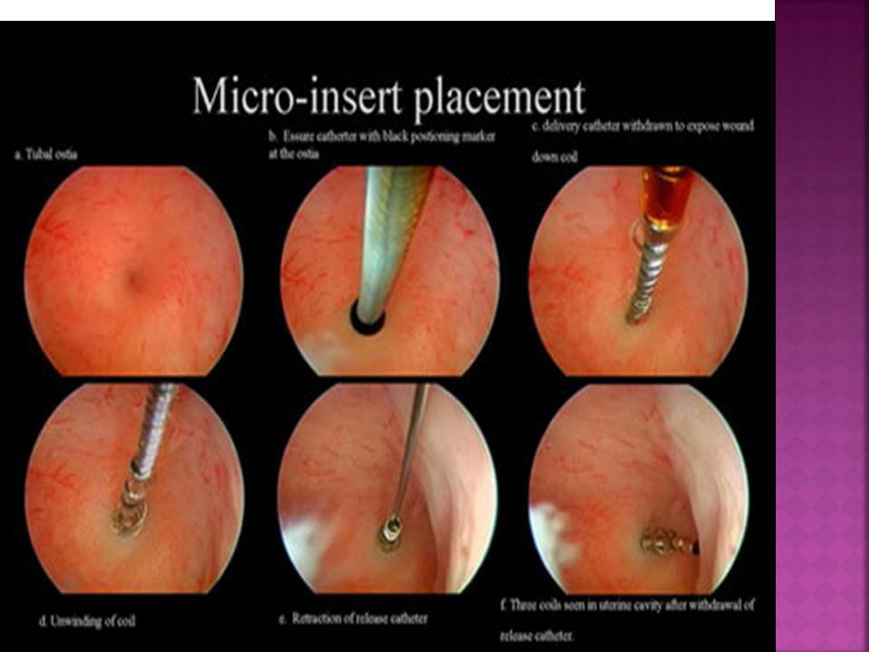
Essure sistem (Flexible micro insert) Sinha D et al. BJOG 2007
Nitinol Dacron intra tubal araç Litta P et al. Human Reprod. 2005

39
Essure Sİstem

41
Essure Sİstem

43
Essure Sİstem Mikro insert 102 kadın çalışmaya dahil edildi.
100 hastaya başarı ile proksimal tubaya mikroinsert yerleştirilmiştir. (% 98) Kerin JF et al. J am Assoc Gynecol Laparoscop 2004.
 Kerin JF et al. J am Assoc Gynecol Laparoscop")
44
Essure Sİstem H/S Mikro insert yerleştirilmesi sonrası 2 nci yılda gebelik olgusu Sterilizasyon sonrası HSG ile tubal oklüzyon değerlendirilmemiştir. Postpartum HSG her iki mikroinsert in fundus ve myometriuma gömülü olduğunu ortaya koymuştur. Hastings- Tolsmam et al. Obstet Gynecol 2007

45
Nitinol Dacron İntratubal Sİstem
Prospektif çalışma Anestezi gereksinimi yok 36 hasta, başarı 3 ay sonraki HSG ile değerlendirildi % 88,9 (32/36) bilateral başarılı uygulama % 2,8 (1/36) unilateral başarılı uygulama % 8,3 (3/36) başarısız Anestezi için kontraendikasyon teşkil eden hastalarda tercih edilebilir. Litta P et al. Human Reprod. 2005 Hum Reprod Dec;20(12): Epub 2005 Aug 5. Related Articles, Links Hysteroscopic permanent tubal sterilization using a nitinol-dacron intratubal device without anaesthesia in the outpatient setting: procedure feasibility and effectiveness. Litta P, Cosmi E, Sacco G, Saccardi C, Ciavattini A, Ambrosini G. Department of Gynecological Science and Human Reproduction, University of Padua School of Medicine, Via Giustiniani No. 3, Padua, Italy. BACKGROUND: Hysteroscopic permanent tubal sterilization has recently been introduced, resulting in a non-invasive, safe and effective technique. The aim of this study was to assess the feasibility of outpatient hysteroscopic tubal sterilization using a nitinol-dacron intratubal device without anaesthesia and to assess patient procedure compliance. MATERIALS AND METHODS: We untertook a prospective study of 36 consecutive cases of outpatient hysteroscopic tubal sterilization using a nitinol-dacron intratubal device without anaesthesia. Tubal sterilization was performed by placing the device with the aid of a 5.2-mm continuous-flow operative hysteroscope. At the end of the procedure women were asked to rate the pain experienced on a visual analogue scale (VAS) (0, no discomfort to 100, severe discomfort). Successful device placement was assessed after 3 months by hysterosalpingography and diagnostic hysteroscopy. RESULTS: Successful bilateral placement was obtained in 32 patients (88.9%); in one (2.8%) the placement was monolateral; and in three (8.3%) the procedure failed. Mean operating time was 8.6 +/- 5.3 min. A mean VAS of / was recorded. CONCLUSIONS: The nitinol-dacron intratubal device is safe, appears to be effective long-term, is non-invasive and can be used in the outpatient setting without anaesthesia. Low-level discomfort was experienced by the patients. Limitations of its use include that it is not effective immediately, it is irreversible, it requires special equipment and training, and it is difficult to use in cases of uterine anomalies. We conclude that this method may be offered to all woman asking for permanent tubal sterilization, particularly those who refuse or have contraindications for anaesthesia. PMID: [PubMed - indexed for MEDLINE]
 bilateral başarılı uygulama. % 2,8 (1/36) unilateral başarılı uygulama. % 8,3 (3/36) başarısız. Anestezi için kontraendikasyon teşkil eden hastalarda tercih edilebilir. Litta P et al. Human Reprod Hum Reprod Dec;20(12): Epub 2005 Aug 5. Related Articles, Links. Hysteroscopic permanent tubal sterilization using a nitinol-dacron intratubal device without anaesthesia in the outpatient setting: procedure feasibility and effectiveness. Litta P, Cosmi E, Sacco G, Saccardi C, Ciavattini A, Ambrosini G. Department of Gynecological Science and Human Reproduction, University of Padua School of Medicine, Via Giustiniani No. 3, Padua, Italy. BACKGROUND: Hysteroscopic permanent tubal sterilization has recently been introduced, resulting in a non-invasive, safe and effective technique. The aim of this study was to assess the feasibility of outpatient hysteroscopic tubal sterilization using a nitinol-dacron intratubal device without anaesthesia and to assess patient procedure compliance. MATERIALS AND METHODS: We untertook a prospective study of 36 consecutive cases of outpatient hysteroscopic tubal sterilization using a nitinol-dacron intratubal device without anaesthesia. Tubal sterilization was performed by placing the device with the aid of a 5.2-mm continuous-flow operative hysteroscope. At the end of the procedure women were asked to rate the pain experienced on a visual analogue scale (VAS) (0, no discomfort to 100, severe discomfort). Successful device placement was assessed after 3 months by hysterosalpingography and diagnostic hysteroscopy. RESULTS: Successful bilateral placement was obtained in 32 patients (88.9%); in one (2.8%) the placement was monolateral; and in three (8.3%) the procedure failed. Mean operating time was 8.6 +/- 5.3 min. A mean VAS of / was recorded. CONCLUSIONS: The nitinol-dacron intratubal device is safe, appears to be effective long-term, is non-invasive and can be used in the outpatient setting without anaesthesia. Low-level discomfort was experienced by the patients. Limitations of its use include that it is not effective immediately, it is irreversible, it requires special equipment and training, and it is difficult to use in cases of uterine anomalies. We conclude that this method may be offered to all woman asking for permanent tubal sterilization, particularly those who refuse or have contraindications for anaesthesia. PMID: [PubMed - indexed for MEDLINE]")
46
Kataterİzasyon ve Tubal Etkİlerİ
Jinekolojik endikasyonlarla TAH/BSO uygulanan 20 hastadan elde edilen materyale 3 F çaplı embryo transfer katater ile kataterizasyon uygulandı. Kontralateral tüpler kontrol grubu Elektron mikroskopisi ile ultrastruktürel yapılar değerlendirilmiş. Silier ve non silier hücreler arasında anormal dezmozom yüzdesi ve bazal membran yüzdesi arasında anlamlı fark yok. Kitamura S et al. J Assist Reprod Genet 1998 J Assist Reprod Genet Aug;15(7):411-7. Related Articles, Links Ultrastructural evaluation following catheterization of the fallopian tube with a hysteroscopic catheter. Kitamura S, Miyazaki T, Iwata S, Akaboshi K, Osawa Y, Yoshimura Y. Department of Obstetrics and Gynecology, School of Medicine, Keio University, Tokyo, Japan. PURPOSE: Our purpose was to assess the morphology and ultrastructural changes in the tubal epithelium following catheterization of the fallopian tube. METHODS: Fallopian tubes were obtained from 20 women who had undergone hysterectomies. Catheterization was performed in 20 tubes using a catheter developed for hysteroscopic tubal embryo transfer. The catheter has a 3-French diameter, tapering to 2 French (0.66 mm) at the tip portion. The 20 contralateral tubes served as controls and were not catheterized. Ultrastructural changes were examined by scanning electron microscopy and transmission electron microscopy. RESULTS: Scanning electron microscopy showed no transformation or defects of the tubal epithelium surface in catheterized or control tubes. Transmission electron microscopy showed no significant differences in the percentage of abnormal desmosomes and the percentage of basement membrane in ciliated and nonciliated cells between catheterized and noncatheterized tubes. No transformation or defects were observed in catheterized or noncatheterized tubes. CONCLUSIONS: These findings suggest that catheterization of the tube using a hysteroscopic catheter caused no acute damage to the tubal epithelium.
: Related Articles, Links. Ultrastructural evaluation following catheterization of the fallopian tube with a hysteroscopic catheter. Kitamura S, Miyazaki T, Iwata S, Akaboshi K, Osawa Y, Yoshimura Y. Department of Obstetrics and Gynecology, School of Medicine, Keio University, Tokyo, Japan. PURPOSE: Our purpose was to assess the morphology and ultrastructural changes in the tubal epithelium following catheterization of the fallopian tube. METHODS: Fallopian tubes were obtained from 20 women who had undergone hysterectomies. Catheterization was performed in 20 tubes using a catheter developed for hysteroscopic tubal embryo transfer. The catheter has a 3-French diameter, tapering to 2 French (0.66 mm) at the tip portion. The 20 contralateral tubes served as controls and were not catheterized. Ultrastructural changes were examined by scanning electron microscopy and transmission electron microscopy. RESULTS: Scanning electron microscopy showed no transformation or defects of the tubal epithelium surface in catheterized or control tubes. Transmission electron microscopy showed no significant differences in the percentage of abnormal desmosomes and the percentage of basement membrane in ciliated and nonciliated cells between catheterized and noncatheterized tubes. No transformation or defects were observed in catheterized or noncatheterized tubes. CONCLUSIONS: These findings suggest that catheterization of the tube using a hysteroscopic catheter caused no acute damage to the tubal epithelium.")
47
Komplİkasyonlar Kornual Perforasyon Uterin perforasyon
En sık görülen komplikasyondur. Genellikle spontan olarak iyileşir. Uterin perforasyon Nadirdir. Postoperatif endometritis ve salpenjitis

Benzer bir sunumlar


 27.03.2008.>")


 Aşağıdaki şekillerden hangisi karedir? AB C D.>")


