Sunuyu indir
Sunum yükleniyor. Lütfen bekleyiniz

1
Jinekolojik Endokrinolojide Hormonal Değerlendirme
Dr.Engin Oral İstanbul Üniversitesi Cerrahpaşa Tıp Fakültesi Kadın Hastalıkları ve Doğum ABD Reprodüktif Endokrinoloji BilimDalı

2
Jinekolojik Endokrinolojide Patolojiler
Over rezervi PKOS Hiperandrojenemi Hiperprolaktinemi Amenore Menopoz Sonuç

4
Gonadotropinler

5
Lipid panel (cholesterol, HDL, LDL, and triglycerides)
Laboratuar tetkikleri LH FSH TSH Prolactin Lipid panel (cholesterol, HDL, LDL, and triglycerides) Fasting insulin level 2-hour 75-g glucose tolerance test DHEAS Testosterone Free testosterone 17-Hydroxyprogesterone
 Fasting insulin level. 2-hour 75-g glucose tolerance test. DHEAS. Testosterone. Free testosterone. 17-Hydroxyprogesterone.")
7
Cut-off Values for the Most Commonly Used Ovarian Reserve Tests

8
Assessment of ovarian reserve with anti-Müllerian hormone: a comparison of the predictive value of anti-Müllerian hormone, follicle-stimulating hormone, inhibin B, and age OBJECTIVE: The objective of this study was to evaluate basal anti- STUDY DESIGN: Frozen basal menstrual cycle day 3 serum samples treatment. Müllerian hormone as a marker for ovarian responsiveness to fertility were evaluated retrospectively for anti-Müllerian hormone, inhibin B, RESULTS: Anti-Müllerian hormone values correlated the best with the (93 patients) and compared with in vitro fertilization records. and follicle-stimulating hormone levels in 123 in vitro fertilization cycles .01), inhibin B (P .05), luteinizing hormone (P .05), and estradiol 0.323; P .01), follicle-stimulating hormone (r ; P number of retrieved oocytes (r ; P .001) relative to age (r (r0.190; P .05). Receiver operating characteristic curve analysis 0.81; P ) relative to age (r 0.74; P .005), follicle-stimulating anti-Müllerian hormone had the largest area under the curve (AUC demonstrated that, for the prediction of 4 oocytes retrieved, oocytes, anti-Müllerian hormone had the largest area under the curve (0.54; P .05). Similarly, for the prediction of 15 retrieved hormone (0.71; P .02), inhibin B (0.66; P .03), and estradiol (0.80; P ) relative to age (0.63; P .02), follicle-stimulating CONCLUSION: Anti-Müllerian hormone correlates better than age, follicle- (0.58; P .05). hormone (0.64; P .005), inhibin B (r 0.57; P .05), and estradiol curves estimated that anti-Müllerian hormone accurately with the number of retrieved oocytes. Receiver operating characteristic stimulating hormone, luteinizing hormone, inhibin B, and estradiol high sensitivity and specificity. predicts ovarian responsiveness to controlled ovarian stimulation with Ryan M. Riggs, 2008
 and compared with in vitro fertilization records. and follicle-stimulating hormone levels in 123 in vitro fertilization cycles. .01), inhibin B (P .05), luteinizing hormone (P .05), and estradiol ; P .01), follicle-stimulating hormone (r 0.317; P. number of retrieved oocytes (r 0.539; P .001) relative to age (r. (r0.190; P .05). Receiver operating characteristic curve analysis. 0.81; P .0001) relative to age (r 0.74; P .005), follicle-stimulating. anti-Müllerian hormone had the largest area under the curve (AUC. demonstrated that, for the prediction of 4 oocytes retrieved, oocytes, anti-Müllerian hormone had the largest area under the curve. (0.54; P .05). Similarly, for the prediction of 15 retrieved. hormone (0.71; P .02), inhibin B (0.66; P .03), and estradiol. (0.80; P .0001) relative to age (0.63; P .02), follicle-stimulating. CONCLUSION: Anti-Müllerian hormone correlates better than age, follicle- (0.58; P .05). hormone (0.64; P .005), inhibin B (r 0.57; P .05), and estradiol. curves estimated that anti-Müllerian hormone accurately. with the number of retrieved oocytes. Receiver operating characteristic. stimulating hormone, luteinizing hormone, inhibin B, and estradiol. high sensitivity and specificity. predicts ovarian responsiveness to controlled ovarian stimulation with. Ryan M. Riggs,")
9
Over rezervini belirleme endikasyonları
İleri kadın yaşı (>35 yaş ?) Geçirilmiş over cerrahisi Tek over Açıklanamayan infertilite Sigara kullanımı Daha evvelki tedavilerde başarısızlık Ailede erken menopoz hikayesi Kemoterapi, radyoterapi Evre III-IV endometriozis HERKESE
 Geçirilmiş over cerrahisi. Tek over. Açıklanamayan infertilite. Sigara kullanımı. Daha evvelki tedavilerde başarısızlık. Ailede erken menopoz hikayesi. Kemoterapi, radyoterapi. Evre III-IV endometriozis. HERKESE.")
10
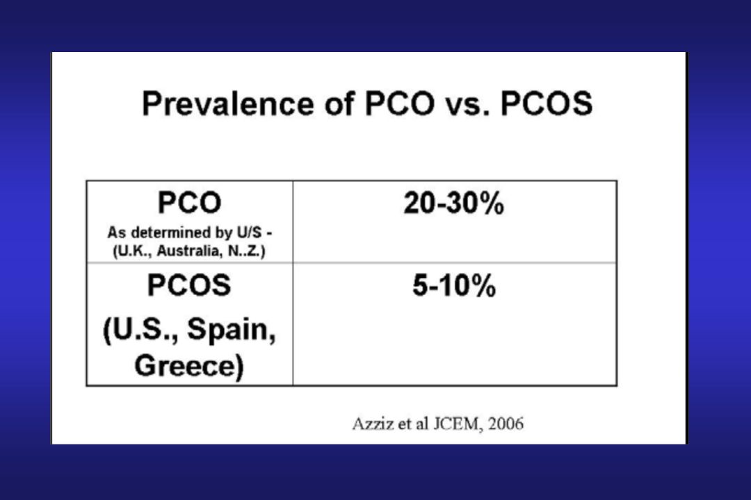
POLYCYSTIC OVARY SYNDROME (PCOS)
Sixty-five to 85% of all women with androgen excess are diagnosed as having PCOS The findings in PCOS are variable, with 40% to 60% of patients obese, 60% to 90% hirsute, 50% to 90% oligoamenorrheic, and 55% to 75% infertile.

11
The Hypothalamic–Pituitary–Ovarian Axis and the Role of Insulin.
Increased ovarian androgen biosynthesis in the polycystic ovary syndrome results from abnormalities at all levels of the hypothalamic–pituitary– ovarian axis. The increased frequency of luteinizing hormone (LH) pulses in the polycystic ovary syndrome appears to result from an increased frequency of hypothalamic gonadotropin-releasing hormone (GnRH) pulses. The latter can result from an intrinsic abnormality in the hypothalamic GnRH pulse generator, favoring the production of luteinizing hormone over follicle-stimulating hormone (FSH) in patients with the polycystic ovary syndrome, in whom the administration of progesterone can restrain the rapid pulse frequency. By whatever mechanism, the relative increase in pituitary secretion of luteinizing hormone leads to an increase in androgen production by ovarian theca cells. Increased efficiency in the conversion of androgenic precursors in theca cells leads to enhanced production of androstenedione, which is then converted by 17 b -hydroxysteroid dehydrogenase (17 ) to form testosterone or aromatized by the aromatase enzyme to form estrone. Within the granulosa cell, estrone is then converted into estradiol by 17 . Numerous autocrine, paracrine, and endocrine factors modulate the effects of both luteinizing hormone and insulin on the androgen production of theca cells; insulin acts synergistically with luteinizing hormone to enhance androgen production. Insulin also inhibits hepatic synthesis of sex hormone–binding globulin, the key circulating protein that binds to testosterone and thus increases the proportion of testosterone that circulates in the unbound, biologically available, or “free,” state. Testosterone inhibits and estrogen stimulates hepatic synthesis of sex hormone–binding globulin. The abbreviation scc denotes side-chain cleavage enzyme, StAR steroidogenic acute regulatory protein, and 3 -HSD 3 -hydroxysteroid dehydrogenase. Solid arrows denote a higher degree of stimulation than dashed arrows. David A. Ehrmann, 2005
 pulses in the polycystic ovary syndrome appears to result from an. increased frequency of hypothalamic gonadotropin-releasing hormone (GnRH) pulses. The latter can result from an intrinsic abnormality in. the hypothalamic GnRH pulse generator, favoring the production of luteinizing hormone over follicle-stimulating hormone (FSH) in patients. with the polycystic ovary syndrome, in whom the administration of progesterone can restrain the rapid pulse frequency. By whatever mechanism, the relative increase in pituitary secretion of luteinizing hormone leads to an increase in androgen production by ovarian theca cells. Increased. efficiency in the conversion of androgenic precursors in theca cells leads to enhanced production of androstenedione, which is then. converted by 17. b. -hydroxysteroid dehydrogenase (17. ) to form testosterone or aromatized by the aromatase enzyme to form estrone. Within. the granulosa cell, estrone is then converted into estradiol by Numerous autocrine, paracrine, and endocrine factors modulate the effects. of both luteinizing hormone and insulin on the androgen production of theca cells; insulin acts synergistically with luteinizing hormone. to enhance androgen production. Insulin also inhibits hepatic synthesis of sex hormone–binding globulin, the key circulating protein that. binds to testosterone and thus increases the proportion of testosterone that circulates in the unbound, biologically available, or free, state. Testosterone inhibits and estrogen stimulates hepatic synthesis of sex hormone–binding globulin. The abbreviation scc denotes side-chain. cleavage enzyme, StAR steroidogenic acute regulatory protein, and 3. -HSD 3. -hydroxysteroid dehydrogenase. Solid arrows denote a higher. degree of stimulation than dashed arrows. David A. Ehrmann,")
13
Criteria for the diagnosis of polycystic ovary syndrome (PCOS)
")
14
Oligo- or anovulation: Ovulation occurs less than once every 35 days.
Hyperandrogenism: Clinical signs include hirsutism, acne, alopecia (male-pattern balding) and frank virilization. Biochemical indicators include raised concentrations of total testosterone and androstendione, and an elevated free androgen index that entails the measurement of total testosterone and sex hormone binding globulin (SHBG). However, the measurement of these biochemical markers for hyperandrogenism has proved markedly inconsistent due to problems with various assays. Polycystic ovaries: The presence of 12 or more follicles in either ovary measuring 2–9 mm in diameter and/or increased ovarian volume (>10 mL).
 and frank virilization. Biochemical indicators include raised concentrations of total testosterone and androstendione, and an elevated free androgen index that entails the measurement of total testosterone and sex hormone binding globulin (SHBG). However, the measurement of these biochemical markers for hyperandrogenism has proved markedly inconsistent due to problems with various assays. Polycystic ovaries: The presence of 12 or more follicles in either ovary measuring 2–9 mm in diameter and/or increased ovarian volume (>10 mL).")
15
Klinik bulgular (%) Menstrüel düzensizlik 66 İnfertilite 75
Hirsutizm 66 Akne 35 Obezite 38 LH Artışı 40 T artışı 30 Hiperinsülinemi Obez Zayıf Homburg R, 2003

16
ANDROGEN EXCESS SOCIETY, 2006

17
Endocrine and metabolic differences among phenotypic expressions of polycystic ovary syndrome according to the 2003 Rotterdam consensus criteria OBJECTIVE: The Rotterdam criteria extend the phenotypic spectrum of polycystic ovary syndrome (PCOS). We characterized endocrine and metabolic differences among women meeting the National Institutes of Health (NIH) definition for PCOS vs two novel phenotypes established by the European Society of Human Reproduction and Embryology/ American Society for Reproductive Medicine definition. STUDY DESIGN: Endocrine and metabolic data from a retrospective analysis of 160 age- and weight-matched women with PCOS and 23 controls were compared. Insulin sensitivity indices were correlated with androgens, gonadotropins, and lipids within each phenotype. RESULTS: Ovarian and adrenal androgens were highest in the NIHdefined PCOS group, lowest in the nonhyperandrogenic PCOS group, and intermediate in the hyperandrogenic ovulatory PCOS population. Insulin sensitivity indices, gonadotropins, and lipids were similar across all PCOS phenotypes. The magnitude of insulin resistance correlated with free testosterone only in the NIH-defined group. CONCLUSION: Androgen levels are the major distinguishing endocrine feature differentiating phenotypic expressions of PCOS. Hyperinsulinemia correlates with free testosterone levels only in traditional NIHdefined women with PCOS. In summary, the data presented suggest that androgen production and secretion is the most prominent endocrine and metabolic factor differentiating phenotypic expressions of PCOS. In addition, insulin appears to play a more prominent role in ovarian androgen production in the NIH phenotype (HA/OD with or without PCO) than in the other subpopulations of PCOS patients. From an endocrine and metabolic standpoint, the OD/PCO phenotype is the mildest expression of the PCOS spectrum. Robert P. Kauffman, 2008
. We characterized endocrine and. metabolic differences among women meeting the National Institutes of. Health (NIH) definition for PCOS vs two novel phenotypes established. by the European Society of Human Reproduction and Embryology/ American Society for Reproductive Medicine definition. STUDY DESIGN: Endocrine and metabolic data from a retrospective. analysis of 160 age- and weight-matched women with PCOS and 23. controls were compared. Insulin sensitivity indices were correlated. with androgens, gonadotropins, and lipids within each phenotype. RESULTS: Ovarian and adrenal androgens were highest in the NIHdefined. PCOS group, lowest in the nonhyperandrogenic PCOS. group, and intermediate in the hyperandrogenic ovulatory PCOS. population. Insulin sensitivity indices, gonadotropins, and lipids. were similar across all PCOS phenotypes. The magnitude of insulin. resistance correlated with free testosterone only in the NIH-defined. group. CONCLUSION: Androgen levels are the major distinguishing endocrine. feature differentiating phenotypic expressions of PCOS. Hyperinsulinemia. correlates with free testosterone levels only in traditional NIHdefined. women with PCOS. In summary, the data presented suggest. that androgen production and secretion. is the most prominent endocrine. and metabolic factor differentiating phenotypic. expressions of PCOS. In addition, insulin appears to play a more. prominent role in ovarian androgen. production in the NIH phenotype. (HA/OD with or without PCO) than in. the other subpopulations of PCOS patients. From an endocrine and metabolic. standpoint, the OD/PCO phenotype is. the mildest expression of the PCOS. spectrum. Robert P. Kauffman,")
18
ANDROGEN EXCESS SOCIETY, 2006

19
Suggested diagnostic evaluation for PCOS
In women with oligomenorrhea, in addition to measurement of serum hCG to rule out pregnancy, minimal laboratory testing should include measurements of serum prolactin, thyrotropin, and FSH to rule out hyperprolactinemia, thyroid disease, and ovarian failure, respectively. (See "Etiology, diagnosis, and treatment of secondary amenorrhea", section on Diagnosis). We do not routinely measure serum androgen concentrations in women with mild hirsutism. However, in women with moderate-to-severe hirsutism, we typically measure a total testosterone concentration, and if there are concerns about a possible androgen-secreting tumor causing the hyperandrogenism, we add serum dehydroepiandrosterone sulfate (DHEA-S). (See "Etiology, diagnosis, and treatment of secondary amenorrhea" and see "Evaluation of women with hirsutism"). Approximately 45 percent of women with PCOS have IGT or type 2 diabetes. A fasting glucose measurement is a less sensitive test for diagnosing IGT and diabetes in women with PCOS. (See "Clinical manifestations of polycystic ovary syndrome in adults", section on Risk of type 2 diabetes). Once the diagnosis of PCOS is made, we perform an OGTT in all patients. When this is not practical, a fasting glucose along with a hemoglobin A1C can be obtained. If either one is abnormal, an OGTT should be performed to distinguish between IGT and diabetes. Given the high prevalence of lipid abnormalities in PCOS, we suggest measuring a fasting lipid profile in all women with the disorder. An elevation in free testosterone is the most sensitive test to establish the presence of hyperandrogenemia. This is because elevated insulin levels (a frequent concomitant of PCOS) and elevated androgen levels both act to inhibit hepatic production of sex hormone-binding globulin (SHBG) [9]. However, commercially available free testosterone assays are often unreliable Other biochemical findings that are often, but not universally present, may include elevation of serum luteinizing hormone (LH) concentrations, normal serum estradiol, and increased serum estrone concentrations. None of these hormones are part of the diagnostic criteria for PCOS and therefore, do not need to be measured. Richard S. Legro, 2007
. We do not routinely measure serum androgen concentrations in women with mild hirsutism. However, in women with moderate-to-severe hirsutism, we typically measure a total testosterone concentration, and if there are concerns about a possible androgen-secreting tumor causing the hyperandrogenism, we add serum dehydroepiandrosterone sulfate (DHEA-S). (See Etiology, diagnosis, and treatment of secondary amenorrhea and see Evaluation of women with hirsutism ). Approximately 45 percent of women with PCOS have IGT or type 2 diabetes. A fasting glucose measurement is a less sensitive test for diagnosing IGT and diabetes in women with PCOS. (See Clinical manifestations of polycystic ovary syndrome in adults , section on Risk of type 2 diabetes). Once the diagnosis of PCOS is made, we perform an OGTT in all patients. When this is not practical, a fasting glucose along with a hemoglobin A1C can be obtained. If either one is abnormal, an OGTT should be performed to distinguish between IGT and diabetes. Given the high prevalence of lipid abnormalities in PCOS, we suggest measuring a fasting lipid profile in all women with the disorder. An elevation in free testosterone is the most sensitive test to establish the presence of hyperandrogenemia. This is because elevated insulin levels (a frequent concomitant of PCOS) and elevated androgen levels both act to inhibit hepatic production of sex hormone-binding globulin (SHBG) [9]. However, commercially available free testosterone assays are often unreliable. Other biochemical findings that are often, but not universally present, may include elevation of serum luteinizing hormone (LH) concentrations, normal serum estradiol, and increased serum estrone concentrations. None of these hormones are part of the diagnostic criteria for PCOS and therefore, do not need to be measured. Richard S. Legro,")
20
Ovarian hyperthecosis
COC ile yanıt alınamayan olgular CC ile başarısız indüksiyonlu hiperandrojenik olgular Virilizm ile birlikte olan olgular İnsülin rezistansı, AN gibi metabolik semptom veren olgularda, LH ve FSH düzeyleri normal ya da düşüktür Serum T düzeyleri tümör sınırlarındadır Ovarian hyperthecosis

21
ANDROJENLER Testosteron %50 periferik dönüşüm %50 over ve adrenal
%80 SHBG, %19 albumine %1 serbest (kadınlarda) DHT En güçlü T,AD DHT Androstenodiol glukuronid
 DHT. En güçlü. T,AD DHT. Androstenodiol glukuronid.")
22
ANDROJENLER DHEAS,DHEA Zayıf Adrenal kaynaklı Gebelikte E3 prekürsörü
Puberte başında pubik kıllanma diğer androjenlerin prekürsörü AD Aktif değil Over, adrenal kaynaklı T,DHT’ye dönüşür

23
S H B G azaltanlar artıranlar Obesite Androjen fazlalığı
Kortikosteroid Hipotiroidism Cushing sendromu Akromegali Karaciğer hast. Progestogen Hiperinsülinemi Östrojen fazlalığı Oral kontraseptifler Gebelik Hipertiroidism

24
ANDROJEN FAZLALIĞINDA CİLT BULGULARI
Hirsutizm Akne Androjenik alopesi AN

25
Which androgen to measure?
Free T or free T index were felt to be most sensitive methods of hyperandrogenemia Measurement of total T only may not be a sensitive marker of AE A fraction of patients may have DHEAS elevation Routine assessment of androstenedione is not recommended ESHRE/ASRM Consensus 2003

26
Factors that are known to alter serum testosterone concentrations
Physiological factors Pulsatile release during the day Diurnal rhythm: am > pm Menstrual cycle: luteal > follicular Season (no variation in total testosterone free testosterone shows 30% difference): summer > winter Age (years) in women with and without polycystic ovary syndrome (PCOS): 20s > 40s Analytical factors Cross reactivity with other endogenous steroids Interference by endogenous antibodies Poor performance in the female range: < 8 nmol/l TT concentrations in plasma vary over 3 orders of magnitude depending on age, gender, and the presence of disease. Only 1–3% of T is not bound to plasma proteins, raising questions about whether TT or free T (FT) is the most clinically useful measure. Age- and gender-corrected normal ranges, using a standardized assay, are generally lacking. There is no universally recognized T-calibrating standard.
: summer > winter. Age (years) in women with and without polycystic ovary syndrome (PCOS): 20s > 40s. Analytical factors. Cross reactivity with other endogenous steroids. Interference by endogenous antibodies. Poor performance in the female range: < 8 nmol/l. TT concentrations in plasma vary over 3 orders of magnitude depending on age, gender, and the presence of disease. Only 1–3% of T is not bound to plasma proteins, raising questions about whether TT or free T (FT) is the most clinically useful measure. Age- and gender-corrected normal ranges, using a standardized assay, are generally lacking. There is no universally recognized T-calibrating standard.")
27
Causes of hirsutism. • Polycystic ovary syndrome • Idiopathic
• Late-onset congenital adrenal hyperplasia • Cushing's syndrome ° Cushing's disease (ACTH-secreting pituitary tumour) ° Ectopic ACTH secretion by non-pituitary tumour (bronchus Table, thyroid) ° Autonomous cortisol secretion by adrenal or ovarian tumour ° Ectopic corticotrophin secretion by tumour (very rare) • Androgen-secreting tumours of the ovary ° Sex-cord stromal cell tumours ° Adrenal-like tumours of the ovary • Androgen-secreting tumours of the adrenal ° Adenomas ° Adenocarcinomas • Iatrogenic ° Testosterone ° Danazol ° Glucocorticoids
 ° Ectopic ACTH secretion by non-pituitary tumour (bronchus Table, thyroid) ° Autonomous cortisol secretion by adrenal or ovarian tumour. ° Ectopic corticotrophin secretion by tumour (very rare) • Androgen-secreting tumours of the ovary. ° Sex-cord stromal cell tumours. ° Adrenal-like tumours of the ovary. • Androgen-secreting tumours of the adrenal. ° Adenomas. ° Adenocarcinomas. • Iatrogenic. ° Testosterone. ° Danazol. ° Glucocorticoids.")
28
Androgen Excess in Women: Experience with Over 1000 Consecutive Patients
R. AZZIZ, 2004

29
Akantozis Nigricans Acanthosis nigricans and hirsutism in a 29-year-old patient with polycystic ovary Syndrome Acanthosis nigricans and skin tags in a 37-year-old Hispanic woman with polycystic ovary syndrome. Acanthosis nigricans, a velvety, hyperpigmentation in skin creases arising from the mitogenic action of insulin on the basal cells of the epidermis, is found in the vast majority of these patients Some patients with PCOS also demonstrate acanthosis nigricans. However, it is important that this syndrome not be confused with PCOS and its milder degree of insulin resistance, since patients with the HAIRAN syndrome tend to have a greater degree of associated morbidity, including NIDDM, hypertension, and CVD androgen-secretingneoplasm Patients with the HAIRAN syndrome will generally have basal insulin greater than 80 U/mL if fasting, or above 500 U/mL, after glucose administration

30
Hyperthecosis previously considered hyperthecosis to be a variant of PCOS, it should be noted that the term hyperthecosis simply refers to the histopathologic finding of islands of hyperplastic theca cells located between collections of small atretic follicles (i.e., “cysts”). Most women with hyperthecosis demonstrate high circulating androgen levels, and consequently lower circulating LH and FSH levels (4–8 mIU/mL) Androgen-secreting tumor. Over stromasında luteinize teka hücrelerinin bulunmasıdır. Uzun süreli anovülasyon ve amenore. Yavaş fakat giderek artan virilizasyon. T tümör düzeyinde, DHEAS normal. LH/FSH=1/1 HAİR-AN sık görülür. Tanı histolojik olarak konur. Fertilizasyon şansı çok azdır.
. Most women with hyperthecosis demonstrate high circulating androgen levels, and consequently lower circulating LH and FSH levels (4–8 mIU/mL) Androgen-secreting tumor. Over stromasında luteinize teka hücrelerinin bulunmasıdır. Uzun süreli anovülasyon ve amenore. Yavaş fakat giderek artan virilizasyon. T tümör düzeyinde, DHEAS normal. LH/FSH=1/1. HAİR-AN sık görülür. Tanı histolojik olarak konur. Fertilizasyon şansı çok azdır.")
31
Non-Classic Adrenal Hyperplasia (NCAH)
1% to 5% of hyperandrogenic women are deficient in the activity of adrenal enzymes, particularly 21-hydroxylase (21-OHase) autosomal recessive 17-hydroxyprogesterone (17-HP) hirsutism, acne, and oligo- and/or amenorrhea
 autosomal recessive. 17-hydroxyprogesterone (17-HP) hirsutism, acne, and oligo- and/or amenorrhea.")
32
ACTH Stimülasyon Testi
AD 3-7 günleri Sabah saat 0.25 mg sentetik ACTH (Cortrosyn) IV * IM yapılmamalı Başlangıçta ve 1 saat sonra kan
 IV. * IM yapılmamalı. Başlangıçta ve 1 saat sonra kan.")
33
Serum 17-OHP seviyesi (0.1 –0.8 ng/ml )
LOKAH (-) LOKAH (+) ACTH Stimülasyon Testi ACTH Stimülasyon Testi Gereksiz < 10 ng/ml > 10 ng/ml Heterozigot LOKAH LOKAH Normal
 LOKAH. (+) ACTH Stimülasyon. Testi. ACTH Stimülasyon. Testi Gereksiz. < 10 ng/ml. > 10 ng/ml. Heterozigot. LOKAH. LOKAH. Normal.")
34
Cushing Syndrome adrenal neoplasm, ectopic ACTH-producing tumor, or pituitary tumor/Cushing disease centripetal fat distribution, thinning of the skin with striae, glucose intolerance, osteoporosis, and proximal muscle weakness menstrual irregularities Cushing syndrome is clinically suspected, adrenal corticoid hyperfunction may be ruled out by oral dexamethasone, 1 mg at 11:00 PM, measuring cortisol at 8:00 AM the following morning. The normal response is a level of less than 5 g/dL. Alternatively, a 24-hour free urinary cortisol can be measured and should be less than 100 g/24 hours. If these measures are elevated, a low-dose suppression (2 mg/ day dexamethasonex two days) should be performed and, if abnormal, the diagnosis of hypercortisolemia (Cushing syndrome) is confirmed.
 should be performed and, if abnormal, the diagnosis of hypercortisolemia (Cushing syndrome) is confirmed.")
35
Androgenic Tumors ovary or adrenal
onset of hyperandrogenism is sudden, and when progression is rapid, or when frank virilization is present Virilizing ovarian tumors, including Sertoli-Leydig cell and lipoid cell tumors, generally exhibit low malignancy potential In young women the possibility of an androgen-secreting tumour should be considered with the following: serum testosterone values above 150 ng/dl ; serum-free testosterone values above 2 ng/dl ; serum dehydroepiandrosterone sulphate values above 700 µg per dl It should be recognized that 20% and 10% of androgen-producing ovarian and adrenal tumors, respectively, may have testosterone levels below this value Androgen-producing tumors can be suspected when the circulating testosterone is persistently greater than 200 ng/dL (HAIRAN syndrome, hyperthecosis, or polycystic ovary syndrome) Adrenal tumors can be suspected when the circulating DHEAS level is persistently greater than 7,000 ng/mL
 Adrenal tumors can be suspected when the circulating DHEAS level is persistently greater than 7,000 ng/mL.")
36
Iatrogenic Causes Exogenous androgens Androgenic steroids Danazol
Glucocorticoids

37
“Idiopathic” Hirsutism
Approximately 15% to 30% of hirsute women do not have ovulatory abnormalities and usually have normal levels of circulating androgens In many of these patients, skin 5-reductase activity is excessive, leading to higher skin concentrations of the active androgen dihydrotestosterone It is important to note that approximately 40% of hirsute women claiming to have “regular menstrual cycles” are actually oligo-ovulatory when evaluated more carefully

38
Hirsutism The Endocrine Society Clinical Practice Guidelines recommend biochemical testing in women with moderate or severe hirsutism, or hirsutism of any degree if it is sudden in onset and rapidly progressive, or associated with irregular menses, obesity, or evidence of virilization (clitoromegaly) The Guidelines suggest first measuring an early morning total testosterone concentration. Although a free testosterone concentration is a more sensitive indicator of androgen excess, most available assays are inaccurate Another approach that many clinicians use, is initial measurement of serum testosterone, prolactin, and DHEA-S, followed by additional testing when indicated Martin, KA, Chang, RJ, Ehrmann, DA, et al. Evaluation and treatment of hirsutism in premenopausal women: an endocrine society clinical practice guideline. J Clin Endocrinol Metab 2008; 93:1105 Martin KA, 2008
 The Guidelines suggest first measuring an early morning total testosterone concentration. Although a free testosterone concentration is a more sensitive indicator of androgen excess, most available assays are inaccurate. Another approach that many clinicians use, is initial measurement of serum testosterone, prolactin, and DHEA-S, followed by additional testing when indicated. Martin, KA, Chang, RJ, Ehrmann, DA, et al. Evaluation and treatment of hirsutism in premenopausal women: an endocrine society clinical practice guideline. J Clin Endocrinol Metab 2008; 93:1105. Martin KA,")
39
Differential diagnosis
ANDROGEN EXCESS SOCIETY, 2006

40
Prolaktin Prolaktin hipotalamustan dopaminin inhibitör kontrolü altında Otonom hipersekresyon, pulsatil GnRH sekresyonunu bozar. Hiperprolaktinemi Fizyolojik (< 50 ng/mL): gebelik, laktasyon, uyku, yoğun egzersiz, stres, cinsellik, yemek
: gebelik, laktasyon, uyku, yoğun egzersiz, stres, cinsellik, yemek.")
41
Causes of hyperprolactinaemia

42
PRL hormon biosentezi Orijinal matür PRL RNA sı 227 AA den oluşan sekansı kodlar Üretim sonrası molekül şu etkilere maruz kalır : Degradasyon Polimerizasyon Glikozilasyon (PRL etkinliğinin devamında gereklidir) Fosforilasyon Bu etkiler sonucu oluşan moleküllerin bioaktiviteleri farklıdır Polimerizasyon oranında bioaktivite düşer (MakroPRL) Monomerik % 80-90 Dimerik % 8-20 Polimerik % 1-5
 Fosforilasyon. Bu etkiler sonucu oluşan moleküllerin bioaktiviteleri farklıdır. Polimerizasyon oranında bioaktivite düşer (MakroPRL) Monomerik % Dimerik % Polimerik % 1-5.")
43
Macroprolactin PRL may form immune complexes, generally with an immunoglobulin G antibody, to produce a biologically inactive form called ‘macroprolactin’, which has a molecular mass of more than 150 kDa. This is registered by most PRL immunoassays and hence serum PRL levels are reported to be high. Since misdiagnosis of hyperprolactinaemia due to the presence of macroprolactin may lead to patient mismanagement, this possibility should be considered in cases with no apparent hyperprolactinaemic symptoms. Polyethylene glycol precipitation is the method of choice to confirm macroprolactinaemia, which in itself has no clinical significance, although it should be remembered that genuine pituitary pathology may co-exist in nearly 5% of such cases. Two causes of hyperprolactinemia due to decreased clearance of prolactin include chronic renal failure (discussed above) and big prolactin. The most common form of prolactin in serum is 23 kD in size and is not glycosylated, but a small amount of a 25 kD glycosylated form can also be detected. In rare cases, glycosylated prolactin, which appears to circulate in aggregates, accounts for most of the prolactin [41]. In this situation the prolactin has been called "big prolactin" and the condition referred to as "macroprolactinemia." The elevated serum prolactin concentration in these patients can be distinguished from hyperprolactinemia of other causes by gel filtration or polyethylene glycol precipitation [42]. In one series of 1106 patients with hyperprolactinemia, approximately 10 percent had macroprolactinemia [43]. The clinical manifestations of macroprolactinemia were described in a series of 55 women ages 18 to 55. None had a history of amenorrhea, eight had oligomenorrhea before age 40, and one had galactorrhea [44]. All subjects had pituitary imaging; no macroadenomas and four microadenomas were seen (consistent with the prevalence of incidentalomas in the normal population). Similar results were seen in a second study of 51 patients [45]. Thus, macroprolactinemia appears to be a benign clinical condition [44]. Equally rare is a raised serum prolactin level due to complexing of normal-sized prolactin with circulating prolactin antibodies [46]. In this situation, the free prolactin concentration is normal and causes no biologic abnormalities. These entities are not of clinical significance directly, but are of clinical significance in an indirect way, because they can be misdiagnosed, and treated, as ordinary hyperprolactinemia [47]. Misdiagnosis can be avoided by asking the laboratory to pretreat the serum with polyethylene glycol to precipitate the macroprolactin before the immunoassay for prolactin.
 and big prolactin. The most common form of prolactin in serum is 23 kD in size and is not glycosylated, but a small amount of a 25 kD glycosylated form can also be detected. In rare cases, glycosylated prolactin, which appears to circulate in aggregates, accounts for most of the prolactin [41]. In this situation the prolactin has been called big prolactin and the condition referred to as macroprolactinemia. The elevated serum prolactin concentration in these patients can be distinguished from hyperprolactinemia of other causes by gel filtration or polyethylene glycol precipitation [42]. In one series of 1106 patients with hyperprolactinemia, approximately 10 percent had macroprolactinemia [43]. The clinical manifestations of macroprolactinemia were described in a series of 55 women ages 18 to 55. None had a history of amenorrhea, eight had oligomenorrhea before age 40, and one had galactorrhea [44]. All subjects had pituitary imaging; no macroadenomas and four microadenomas were seen (consistent with the prevalence of incidentalomas in the normal population). Similar results were seen in a second study of 51 patients [45]. Thus, macroprolactinemia appears to be a benign clinical condition [44]. Equally rare is a raised serum prolactin level due to complexing of normal-sized prolactin with circulating prolactin antibodies [46]. In this situation, the free prolactin concentration is normal and causes no biologic abnormalities. These entities are not of clinical significance directly, but are of clinical significance in an indirect way, because they can be misdiagnosed, and treated, as ordinary hyperprolactinemia [47]. Misdiagnosis can be avoided by asking the laboratory to pretreat the serum with polyethylene glycol to precipitate the macroprolactin before the immunoassay for prolactin.")
44
James Gibney, 2005 Table 1 summarizes
the reported prevalence of macroprolactinaemia in studies in which all hyperprolactinaemic samples were screened for macroprolactin.

45
Hiperprolaktinemi PRL ölçümleri stressiz bir zamanda, sabah aç olarak yapılmalıdır Testten hemen önce: Göğüs muayenesi Koitus Pelvik muayene Egzersiz yapılmamalı Çok yüksek PRL düzeyleri immunoassay de yanlış negatif sonuç verebileceği için (hook effect – kanca etkisi) makroadenom takiplerinde 1/100 dilüsyon ile PRL ikinci kez tekrar edilmelidir Fizyolojik serum prolaktin düzeyleri 50 ng/ml’in altındadır. Serum prolaktin düzeyleri “two site” immünradyometrik assayler (IRMA) ve kemiluminometrik assayler ( ICMA) ile yüksek duyarlılık ve doğruluk ile ölçülmektedir. Fakat çok büyük prolaktinoması ve çok yüksek serum prolaktin düzeyi olan hastalarda, antikorların satürasyonu prolaktin-antikor sandviçinin oluşumuna engel olabilir. Böylece işaretli antikor düzeyi azalacağından serum prolaktin düzeyi yanlış olarak düşük rapor edilebilir. Kanca etkisi ( hook effect) olarak tanımlanan bu yanılmayı ortadan kaldırmak için makroadenomu olan olgularda prolaktin düzeyleri 1/100 dilüe edilmiş serumda tekrar çalışılmalıdır. Caution should be exercised in interpreting serum prolactin concentrations between 20 and 200 ng/mL (20 to 200 mcg/L SI units) in the presence of a macroadenoma, because of possible artefactually low values due to the "hook effect" [13-15]. This effect occurs when a very high serum prolactin, eg, 5000 ng/mL (5000 mcg/L SI units), saturates both the capture and signal antibodies used in immunoradiometric and chemiluminescent assays, preventing the binding of the two in a "sandwich." The result is an apparent prolactin concentration that is only modestly elevated, suggesting that the macroadenoma is clinically nonfunctioning. The artefact can be avoided by repeating the assay using a 1:100 dilution of serum
 makroadenom takiplerinde 1/100 dilüsyon ile PRL ikinci kez tekrar edilmelidir. Fizyolojik serum prolaktin düzeyleri 50 ng/ml’in altındadır. Serum prolaktin düzeyleri two site immünradyometrik assayler (IRMA) ve kemiluminometrik assayler ( ICMA) ile yüksek duyarlılık ve doğruluk ile ölçülmektedir. Fakat çok büyük prolaktinoması ve çok yüksek serum prolaktin düzeyi olan hastalarda, antikorların satürasyonu prolaktin-antikor sandviçinin oluşumuna engel olabilir. Böylece işaretli antikor düzeyi azalacağından serum prolaktin düzeyi yanlış olarak düşük rapor edilebilir. Kanca etkisi ( hook effect) olarak tanımlanan bu yanılmayı ortadan kaldırmak için makroadenomu olan olgularda prolaktin düzeyleri 1/100 dilüe edilmiş serumda tekrar çalışılmalıdır. Caution should be exercised in interpreting serum prolactin concentrations between 20 and 200 ng/mL (20 to 200 mcg/L SI units) in the presence of a macroadenoma, because of possible artefactually low values due to the hook effect [13-15]. This effect occurs when a very high serum prolactin, eg, 5000 ng/mL (5000 mcg/L SI units), saturates both the capture and signal antibodies used in immunoradiometric and chemiluminescent assays, preventing the binding of the two in a sandwich. The result is an apparent prolactin concentration that is only modestly elevated, suggesting that the macroadenoma is clinically nonfunctioning. The artefact can be avoided by repeating the assay using a 1:100 dilution of serum.")
46
Common causes of primary amenorrhea
Bachmann G,1982; Reindollar RH, 1986

47
The initial useful laboratory tests are FSH, TSH, and
Suggested flow diagram aiding in the evaluation of women with amenorrhea. The initial useful laboratory tests are FSH, TSH, and prolactin. The Practice Committee of the American Society for Reproductive Medicine 2008

48
Common causes of secondary amenorrhea
Reindollar RM, 1981

49
STRAW reproductive aging system
Length decreases -2 days

50
Physiology: perimenopause
Variable hormone levels Estrogen and progesterone levels fluctuate erratically Very high serum estrogen levels may result Slight decline in testosterone with age Santoro et al. J Clin Endocrinol Metab 2000. Burger et al. J Clin Endocrinol Metab 2000.

51
Hyperestrogenism in perimenopause
Santoro et al. J Clin Endocrinol Metab 1996.

52
The Menopausal Transition A Committee Opinion Revised January 2008
In perimenopausal women, estradiol production fluctuates with FSH levels and can reach higher concentrations than those observed in young women under age. Estradiol levels generally do not decrease significantly until late in the MT Despite continuing regular cyclic menstruation, progesterone levels during the early MT are lower than in women of mid-reproductive age and vary inversely with body mass index Testosterone levels do not vary appreciably during the MT a decrease in secretion of inhibin A and inhibin B, and a corresponding increase in activin production may favor increased FSH secretion in the absence of any decrease (and perhaps an increase) in estradiol production. Diagnosis of the MT is based on clinical signs and symptoms. Although hormonal changes occur during the MT, hormone measurements are not useful for predicting the stage of MT or the final menstrual period. Practice Committee of the American Society for Reproductive Medicine
 in estradiol production. Diagnosis of the MT is based on clinical signs and symptoms. Although hormonal changes occur during the MT, hormone measurements are not useful for predicting the stage of MT or the final menstrual period. Practice Committee of the American. Society for Reproductive Medicine.")
Benzer bir sunumlar