Sunuyu indir
Sunum yükleniyor. Lütfen bekleyiniz

1
Sinyal Transdüksiyonun I
Hücreler Arası İletişim 1 Sinyal İletimi 1 Yrd. Doç. Dr. İzzet YELKOVAN SİVAS- 2005

2
External sinyal iletimi: Hedef hücrelere sinyal iletimi
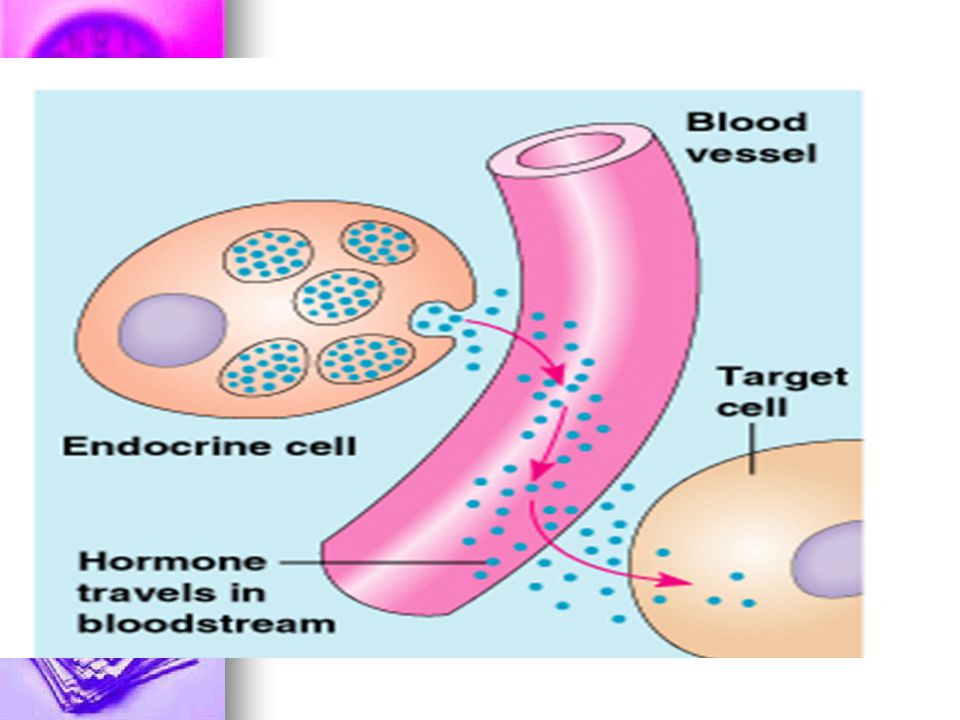
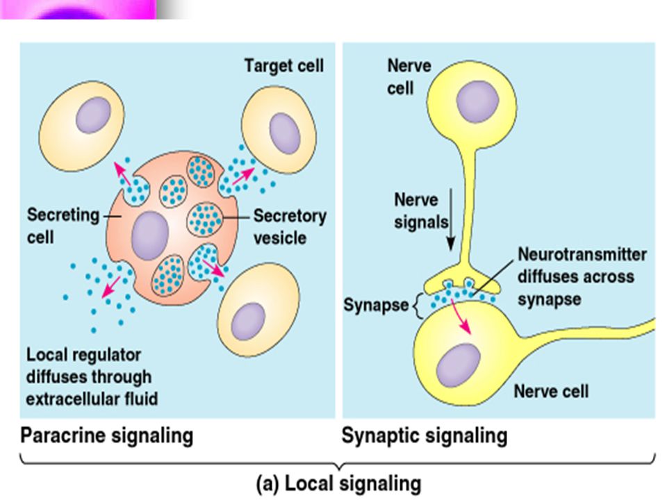
(1) Endokrine sinyal iletimi: sinyal molekülleri (hormonlar) endokrin organ hücrelerince sentezlenirler – sentezlendikleri yerin uzağında başka bir mikroçevredeki hedef hücrelere etki ederler. (2) Parakrin sinyal iletimi: sinyal molekülleri aynı mikroçevreyi paylaşan bir hücre veya hücre grubu tarafında salınır ve yakın çevredeki diğer hücreleri etkiler (Neurotransmitterler ve neurohormonlar). (3) Autokrin sinyal iletimi: sinyal molekülleri bir hücre veya hücre grubundan salınır ve sadece kendileri yanıt oluştururlar (bir çok büyüme faktörü). (4) Hücre – hücre ve hücre matriks arası etkileşimlerde işe karışan moleküller.
 Endokrine sinyal iletimi: sinyal molekülleri (hormonlar) endokrin organ hücrelerince sentezlenirler – sentezlendikleri yerin uzağında başka bir mikroçevredeki hedef hücrelere etki ederler. (2) Parakrin sinyal iletimi: sinyal molekülleri aynı mikroçevreyi paylaşan bir hücre veya hücre grubu tarafında salınır ve yakın çevredeki diğer hücreleri etkiler (Neurotransmitterler ve neurohormonlar). (3) Autokrin sinyal iletimi: sinyal molekülleri bir hücre veya hücre grubundan salınır ve sadece kendileri yanıt oluştururlar (bir çok büyüme faktörü). (4) Hücre – hücre ve hücre matriks arası etkileşimlerde işe karışan moleküller.")
3
External sinyal iletimi: Hedef hücrelere sinyal iletimi
(1) Endokrine sinyal iletimi: sinyal molekülleri (hormonlar) endokrin organ hücrelerince sentezlenirler – sentezlendikleri yerin uzağında başka bir mikroçevredeki hedef hücrelere etki ederler. (2) Parakrin sinyal iletimi: sinyal molekülleri aynı mikroçevreyi paylaşan bir hücre veya hücre grubu tarafında salınır ve yakın çevredeki diğer hücreleri etkiler (Neurotransmitterler ve neurohormonlar). (3) Autokrin sinyal iletimi: sinyal molekülleri bir hücre veya hücre grubundan salınır ve sadece kendileri yanıt oluştururlar (bir çok büyüme faktörü). (4) Hücre – hücre ve hücre matriks arası etkileşimlerde işe karışan moleküller.
 Endokrine sinyal iletimi: sinyal molekülleri (hormonlar) endokrin organ hücrelerince sentezlenirler – sentezlendikleri yerin uzağında başka bir mikroçevredeki hedef hücrelere etki ederler. (2) Parakrin sinyal iletimi: sinyal molekülleri aynı mikroçevreyi paylaşan bir hücre veya hücre grubu tarafında salınır ve yakın çevredeki diğer hücreleri etkiler (Neurotransmitterler ve neurohormonlar). (3) Autokrin sinyal iletimi: sinyal molekülleri bir hücre veya hücre grubundan salınır ve sadece kendileri yanıt oluştururlar (bir çok büyüme faktörü). (4) Hücre – hücre ve hücre matriks arası etkileşimlerde işe karışan moleküller.")
4
External sinyal iletimi: Hedef hücrelere sinyal iletimi
(1) Endokrine sinyal iletimi: sinyal molekülleri (hormonlar) endokrin organ hücrelerince sentezlenirler – sentezlendikleri yerin uzağında başka bir mikroçevredeki hedef hücrelere etki ederler. (2) Parakrin sinyal iletimi: sinyal molekülleri aynı mikroçevreyi paylaşan bir hücre veya hücre grubu tarafında salınır ve yakın çevredeki diğer hücreleri etkiler (Neurotransmitterler ve neurohormonlar). (3) Autokrin sinyal iletimi: sinyal molekülleri bir hücre veya hücre grubundan salınır ve sadece kendileri yanıt oluştururlar (bir çok büyüme faktörü). (4) Hücre – hücre ve hücre matriks arası etkileşimlerde işe karışan moleküller.
 Endokrine sinyal iletimi: sinyal molekülleri (hormonlar) endokrin organ hücrelerince sentezlenirler – sentezlendikleri yerin uzağında başka bir mikroçevredeki hedef hücrelere etki ederler. (2) Parakrin sinyal iletimi: sinyal molekülleri aynı mikroçevreyi paylaşan bir hücre veya hücre grubu tarafında salınır ve yakın çevredeki diğer hücreleri etkiler (Neurotransmitterler ve neurohormonlar). (3) Autokrin sinyal iletimi: sinyal molekülleri bir hücre veya hücre grubundan salınır ve sadece kendileri yanıt oluştururlar (bir çok büyüme faktörü). (4) Hücre – hücre ve hücre matriks arası etkileşimlerde işe karışan moleküller.")
5
External sinyal iletimi: Hedef hücrelere sinyal iletimi
(1) Endokrine sinyal iletimi: sinyal molekülleri (hormonlar) endokrin organ hücrelerince sentezlenirler – sentezlendikleri yerin uzağında başka bir mikroçevredeki hedef hücrelere etki ederler. (2) Parakrin sinyal iletimi: sinyal molekülleri aynı mikroçevreyi paylaşan bir hücre veya hücre grubu tarafında salınır ve yakın çevredeki diğer hücreleri etkiler (Neurotransmitterler ve neurohormonlar). (3) Autokrin sinyal iletimi: sinyal molekülleri bir hücre veya hücre grubundan salınır ve sadece kendileri yanıt oluştururlar (bir çok büyüme faktörü). (4) Hücre – hücre ve hücre matriks arası etkileşimlerde işe karışan moleküller.
 Endokrine sinyal iletimi: sinyal molekülleri (hormonlar) endokrin organ hücrelerince sentezlenirler – sentezlendikleri yerin uzağında başka bir mikroçevredeki hedef hücrelere etki ederler. (2) Parakrin sinyal iletimi: sinyal molekülleri aynı mikroçevreyi paylaşan bir hücre veya hücre grubu tarafında salınır ve yakın çevredeki diğer hücreleri etkiler (Neurotransmitterler ve neurohormonlar). (3) Autokrin sinyal iletimi: sinyal molekülleri bir hücre veya hücre grubundan salınır ve sadece kendileri yanıt oluştururlar (bir çok büyüme faktörü). (4) Hücre – hücre ve hücre matriks arası etkileşimlerde işe karışan moleküller.")
6
External sinyal iletimi: Hedef hücrelere sinyal iletimi
(1) Endokrine sinyal iletimi: sinyal molekülleri (hormonlar) endokrin organ hücrelerince sentezlenirler – sentezlendikleri yerin uzağında başka bir mikroçevredeki hedef hücrelere etki ederler. (2) Parakrin sinyal iletimi: sinyal molekülleri aynı mikroçevreyi paylaşan bir hücre veya hücre grubu tarafında salınır ve yakın çevredeki diğer hücreleri etkiler (Neurotransmitterler ve neurohormonlar). (3) Autokrin sinyal iletimi: sinyal molekülleri bir hücre veya hücre grubundan salınır ve sadece kendileri yanıt oluştururlar (bir çok büyüme faktörü). (4) Hücre – hücre ve hücre matriks arası etkileşimlerde işe karışan moleküller.
 Endokrine sinyal iletimi: sinyal molekülleri (hormonlar) endokrin organ hücrelerince sentezlenirler – sentezlendikleri yerin uzağında başka bir mikroçevredeki hedef hücrelere etki ederler. (2) Parakrin sinyal iletimi: sinyal molekülleri aynı mikroçevreyi paylaşan bir hücre veya hücre grubu tarafında salınır ve yakın çevredeki diğer hücreleri etkiler (Neurotransmitterler ve neurohormonlar). (3) Autokrin sinyal iletimi: sinyal molekülleri bir hücre veya hücre grubundan salınır ve sadece kendileri yanıt oluştururlar (bir çok büyüme faktörü). (4) Hücre – hücre ve hücre matriks arası etkileşimlerde işe karışan moleküller.")
8
External sinyal iletimi: Hedef hücrelere sinyal iletimi
(1) Endokrine sinyal iletimi: sinyal molekülleri (hormonlar) endokrin organ hücrelerince sentezlenirler – sentezlendikleri yerin uzağında başka bir mikroçevredeki hedef hücrelere etki ederler. (2) Parakrin sinyal iletimi: sinyal molekülleri aynı mikroçevreyi paylaşan bir hücre veya hücre grubu tarafında salınır ve yakın çevredeki diğer hücreleri etkiler (Neurotransmitterler ve neurohormonlar). (3) Autokrin sinyal iletimi: sinyal molekülleri bir hücre veya hücre grubundan salınır ve sadece kendileri yanıt oluştururlar (bir çok büyüme faktörü). (4) Hücre – hücre ve hücre matriks arası etkileşimlerde işe karışan moleküller.
 Endokrine sinyal iletimi: sinyal molekülleri (hormonlar) endokrin organ hücrelerince sentezlenirler – sentezlendikleri yerin uzağında başka bir mikroçevredeki hedef hücrelere etki ederler. (2) Parakrin sinyal iletimi: sinyal molekülleri aynı mikroçevreyi paylaşan bir hücre veya hücre grubu tarafında salınır ve yakın çevredeki diğer hücreleri etkiler (Neurotransmitterler ve neurohormonlar). (3) Autokrin sinyal iletimi: sinyal molekülleri bir hücre veya hücre grubundan salınır ve sadece kendileri yanıt oluştururlar (bir çok büyüme faktörü). (4) Hücre – hücre ve hücre matriks arası etkileşimlerde işe karışan moleküller.")
9
External sinyal iletimi: Hedef hücrelere sinyal iletimi
(1) Endokrine sinyal iletimi: sinyal molekülleri (hormonlar) endokrin organ hücrelerince sentezlenirler – sentezlendikleri yerin uzağında başka bir mikroçevredeki hedef hücrelere etki ederler. (2) Parakrin sinyal iletimi: sinyal molekülleri aynı mikroçevreyi paylaşan bir hücre veya hücre grubu tarafında salınır ve yakın çevredeki diğer hücreleri etkiler (Neurotransmitterler ve neurohormonlar). (3) Autokrin sinyal iletimi: sinyal molekülleri bir hücre veya hücre grubundan salınır ve sadece kendileri yanıt oluştururlar (bir çok büyüme faktörü). (4) Hücre – hücre ve hücre matriks arası etkileşimlerde işe karışan moleküller.
 Endokrine sinyal iletimi: sinyal molekülleri (hormonlar) endokrin organ hücrelerince sentezlenirler – sentezlendikleri yerin uzağında başka bir mikroçevredeki hedef hücrelere etki ederler. (2) Parakrin sinyal iletimi: sinyal molekülleri aynı mikroçevreyi paylaşan bir hücre veya hücre grubu tarafında salınır ve yakın çevredeki diğer hücreleri etkiler (Neurotransmitterler ve neurohormonlar). (3) Autokrin sinyal iletimi: sinyal molekülleri bir hücre veya hücre grubundan salınır ve sadece kendileri yanıt oluştururlar (bir çok büyüme faktörü). (4) Hücre – hücre ve hücre matriks arası etkileşimlerde işe karışan moleküller.")
10
External sinyal iletimi: Hedef hücrelere sinyal iletimi
(1) Endokrine sinyal iletimi: sinyal molekülleri (hormonlar) endokrin organ hücrelerince sentezlenirler – sentezlendikleri yerin uzağında başka bir mikroçevredeki hedef hücrelere etki ederler. (2) Parakrin sinyal iletimi: sinyal molekülleri aynı mikroçevreyi paylaşan bir hücre veya hücre grubu tarafında salınır ve yakın çevredeki diğer hücreleri etkiler (Neurotransmitterler ve neurohormonlar). (3) Autokrin sinyal iletimi: sinyal molekülleri bir hücre veya hücre grubundan salınır ve sadece kendileri yanıt oluştururlar (bir çok büyüme faktörü). (4) Hücre – hücre ve hücre matriks arası etkileşimlerde işe karışan moleküller.
 Endokrine sinyal iletimi: sinyal molekülleri (hormonlar) endokrin organ hücrelerince sentezlenirler – sentezlendikleri yerin uzağında başka bir mikroçevredeki hedef hücrelere etki ederler. (2) Parakrin sinyal iletimi: sinyal molekülleri aynı mikroçevreyi paylaşan bir hücre veya hücre grubu tarafında salınır ve yakın çevredeki diğer hücreleri etkiler (Neurotransmitterler ve neurohormonlar). (3) Autokrin sinyal iletimi: sinyal molekülleri bir hücre veya hücre grubundan salınır ve sadece kendileri yanıt oluştururlar (bir çok büyüme faktörü). (4) Hücre – hücre ve hücre matriks arası etkileşimlerde işe karışan moleküller.")
12
External sinyal iletimi: Hedef hücrelere sinyal iletimi
(1) Endokrine sinyal iletimi: sinyal molekülleri (hormonlar) endokrin organ hücrelerince sentezlenirler – sentezlendikleri yerin uzağında başka bir mikroçevredeki hedef hücrelere etki ederler. (2) Parakrin sinyal iletimi: sinyal molekülleri aynı mikroçevreyi paylaşan bir hücre veya hücre grubu tarafında salınır ve yakın çevredeki diğer hücreleri etkiler (Neurotransmitterler ve neurohormonlar). (3) Autokrin sinyal iletimi: sinyal molekülleri bir hücre veya hücre grubundan salınır ve sadece kendileri yanıt oluştururlar (bir çok büyüme faktörü). (4) Hücre – hücre ve hücre matriks arası etkileşimlerde işe karışan moleküller. In such autocrine signaling a cell secretes signaling molecules that can bind back to its own receptors. During development, for example, once a cell has been directed into a particular path of differentiation, it may begin to secrete autocrine signals that reinforce this developmental decision. Because autocrine signaling is most effective when carried out simultaneously by neighboring cells of the same type, it may be used to encourage groups of identical cells to make the same developmental decisions ( Figure 15-5). Autocrine signaling is not confined to development, however. Eicosanoids are signaling molecules that often act in an autocrine mode in mature mammals. These fatty-acid derivatives are made by cells in all mammalian tissues. They are continuously synthesized in the plasma membrane and released to the cell exterior, where they are rapidly degraded by enzymes in extracellular fluid. Made from precursors (mainly arachidonic acid) that are cleaved from membrane phospholipids by phospholipases ( Figure 15-6), they have a wide variety of biological activities, influencing the contraction of smooth muscle and the aggregation of platelets, for example, and participating in pain and inflammatory responses. When cells are activated by tissue damage or by some types of chemical signals, the rate of eicosanoid synthesis is increased; the resulting increase in the local level of eicosanoid influences both the cells that make it and their immediate neighbors.
 Endokrine sinyal iletimi: sinyal molekülleri (hormonlar) endokrin organ hücrelerince sentezlenirler – sentezlendikleri yerin uzağında başka bir mikroçevredeki hedef hücrelere etki ederler. (2) Parakrin sinyal iletimi: sinyal molekülleri aynı mikroçevreyi paylaşan bir hücre veya hücre grubu tarafında salınır ve yakın çevredeki diğer hücreleri etkiler (Neurotransmitterler ve neurohormonlar). (3) Autokrin sinyal iletimi: sinyal molekülleri bir hücre veya hücre grubundan salınır ve sadece kendileri yanıt oluştururlar (bir çok büyüme faktörü). (4) Hücre – hücre ve hücre matriks arası etkileşimlerde işe karışan moleküller. In such autocrine signaling a cell secretes signaling molecules that can bind back to its own receptors. During development, for example, once a cell has been directed into a particular path of differentiation, it may begin to secrete autocrine signals that reinforce this developmental decision. Because autocrine signaling is most effective when carried out simultaneously by neighboring cells of the same type, it may be used to encourage groups of identical cells to make the same developmental decisions ( Figure 15-5). Autocrine signaling is not confined to development, however. Eicosanoids are signaling molecules that often act in an autocrine mode in mature mammals. These fatty-acid derivatives are made by cells in all mammalian tissues. They are continuously synthesized in the plasma membrane and released to the cell exterior, where they are rapidly degraded by enzymes in extracellular fluid. Made from precursors (mainly arachidonic acid) that are cleaved from membrane phospholipids by phospholipases ( Figure 15-6), they have a wide variety of biological activities, influencing the contraction of smooth muscle and the aggregation of platelets, for example, and participating in pain and inflammatory responses. When cells are activated by tissue damage or by some types of chemical signals, the rate of eicosanoid synthesis is increased; the resulting increase in the local level of eicosanoid influences both the cells that make it and their immediate neighbors.")
13
External sinyal iletimi: Hedef hücrelere sinyal iletimi
(1) Endokrine sinyal iletimi: sinyal molekülleri (hormonlar) endokrin organ hücrelerince sentezlenirler – sentezlendikleri yerin uzağında başka bir mikroçevredeki hedef hücrelere etki ederler. (2) Parakrin sinyal iletimi: sinyal molekülleri aynı mikroçevreyi paylaşan bir hücre veya hücre grubu tarafında salınır ve yakın çevredeki diğer hücreleri etkiler (Neurotransmitterler ve neurohormonlar). (3) Autokrin sinyal iletimi: sinyal molekülleri bir hücre veya hücre grubundan salınır ve sadece kendileri yanıt oluştururlar (bir çok büyüme faktörü). (4) Hücre – hücre ve hücre matriks arası etkileşimlerde işe karışan moleküller.
 Endokrine sinyal iletimi: sinyal molekülleri (hormonlar) endokrin organ hücrelerince sentezlenirler – sentezlendikleri yerin uzağında başka bir mikroçevredeki hedef hücrelere etki ederler. (2) Parakrin sinyal iletimi: sinyal molekülleri aynı mikroçevreyi paylaşan bir hücre veya hücre grubu tarafında salınır ve yakın çevredeki diğer hücreleri etkiler (Neurotransmitterler ve neurohormonlar). (3) Autokrin sinyal iletimi: sinyal molekülleri bir hücre veya hücre grubundan salınır ve sadece kendileri yanıt oluştururlar (bir çok büyüme faktörü). (4) Hücre – hücre ve hücre matriks arası etkileşimlerde işe karışan moleküller.")
14
External sinyal iletimi: Hedef hücrelere sinyal iletimi
(1) Endokrine sinyal iletimi: sinyal molekülleri (hormonlar) endokrin organ hücrelerince sentezlenirler – sentezlendikleri yerin uzağında başka bir mikroçevredeki hedef hücrelere etki ederler. (2) Parakrin sinyal iletimi: sinyal molekülleri aynı mikroçevreyi paylaşan bir hücre veya hücre grubu tarafında salınır ve yakın çevredeki diğer hücreleri etkiler (Neurotransmitterler ve neurohormonlar). (3) Autokrin sinyal iletimi: sinyal molekülleri bir hücre veya hücre grubundan salınır ve sadece kendileri yanıt oluştururlar (bir çok büyüme faktörü). (4) Hücre – hücre ve hücre matriks arası etkileşimlerde işe karışan moleküller.
 Endokrine sinyal iletimi: sinyal molekülleri (hormonlar) endokrin organ hücrelerince sentezlenirler – sentezlendikleri yerin uzağında başka bir mikroçevredeki hedef hücrelere etki ederler. (2) Parakrin sinyal iletimi: sinyal molekülleri aynı mikroçevreyi paylaşan bir hücre veya hücre grubu tarafında salınır ve yakın çevredeki diğer hücreleri etkiler (Neurotransmitterler ve neurohormonlar). (3) Autokrin sinyal iletimi: sinyal molekülleri bir hücre veya hücre grubundan salınır ve sadece kendileri yanıt oluştururlar (bir çok büyüme faktörü). (4) Hücre – hücre ve hücre matriks arası etkileşimlerde işe karışan moleküller.")
15
External sinyal iletimi: Hedef hücrelere sinyal iletimi
(1) Endokrine sinyal iletimi: sinyal molekülleri (hormonlar) endokrin organ hücrelerince sentezlenirler – sentezlendikleri yerin uzağında başka bir mikroçevredeki hedef hücrelere etki ederler. (2) Parakrin sinyal iletimi: sinyal molekülleri aynı mikroçevreyi paylaşan bir hücre veya hücre grubu tarafında salınır ve yakın çevredeki diğer hücreleri etkiler (Neurotransmitterler ve neurohormonlar). (3) Autokrin sinyal iletimi: sinyal molekülleri bir hücre veya hücre grubundan salınır ve sadece kendileri yanıt oluştururlar (bir çok büyüme faktörü). (4) Hücre – hücre ve hücre matriks arası etkileşimlerde işe karışan moleküller.
 Endokrine sinyal iletimi: sinyal molekülleri (hormonlar) endokrin organ hücrelerince sentezlenirler – sentezlendikleri yerin uzağında başka bir mikroçevredeki hedef hücrelere etki ederler. (2) Parakrin sinyal iletimi: sinyal molekülleri aynı mikroçevreyi paylaşan bir hücre veya hücre grubu tarafında salınır ve yakın çevredeki diğer hücreleri etkiler (Neurotransmitterler ve neurohormonlar). (3) Autokrin sinyal iletimi: sinyal molekülleri bir hücre veya hücre grubundan salınır ve sadece kendileri yanıt oluştururlar (bir çok büyüme faktörü). (4) Hücre – hücre ve hücre matriks arası etkileşimlerde işe karışan moleküller.")
16
External sinyal iletimi: Hedef hücrelere sinyal iletimi
(1) Endokrine sinyal iletimi: sinyal molekülleri (hormonlar) endokrin organ hücrelerince sentezlenirler – sentezlendikleri yerin uzağında başka bir mikroçevredeki hedef hücrelere etki ederler. (2) Parakrin sinyal iletimi: sinyal molekülleri aynı mikroçevreyi paylaşan bir hücre veya hücre grubu tarafında salınır ve yakın çevredeki diğer hücreleri etkiler (Neurotransmitterler ve neurohormonlar). (3) Autokrin sinyal iletimi: sinyal molekülleri bir hücre veya hücre grubundan salınır ve sadece kendileri yanıt oluştururlar (bir çok büyüme faktörü). (4) Hücre – hücre ve hücre matriks arası etkileşimlerde işe karışan moleküller.
 Endokrine sinyal iletimi: sinyal molekülleri (hormonlar) endokrin organ hücrelerince sentezlenirler – sentezlendikleri yerin uzağında başka bir mikroçevredeki hedef hücrelere etki ederler. (2) Parakrin sinyal iletimi: sinyal molekülleri aynı mikroçevreyi paylaşan bir hücre veya hücre grubu tarafında salınır ve yakın çevredeki diğer hücreleri etkiler (Neurotransmitterler ve neurohormonlar). (3) Autokrin sinyal iletimi: sinyal molekülleri bir hücre veya hücre grubundan salınır ve sadece kendileri yanıt oluştururlar (bir çok büyüme faktörü). (4) Hücre – hücre ve hücre matriks arası etkileşimlerde işe karışan moleküller.")
17
External sinyal iletimi: Hedef hücrelere sinyal iletimi
(1) Endokrine sinyal iletimi: sinyal molekülleri (hormonlar) endokrin organ hücrelerince sentezlenirler – sentezlendikleri yerin uzağında başka bir mikroçevredeki hedef hücrelere etki ederler. (2) Parakrin sinyal iletimi: sinyal molekülleri aynı mikroçevreyi paylaşan bir hücre veya hücre grubu tarafında salınır ve yakın çevredeki diğer hücreleri etkiler (Neurotransmitterler ve neurohormonlar). (3) Autokrin sinyal iletimi: sinyal molekülleri bir hücre veya hücre grubundan salınır ve sadece kendileri yanıt oluştururlar (bir çok büyüme faktörü). (4) Hücre – hücre ve hücre matriks arası etkileşimlerde işe karışan moleküller.
 Endokrine sinyal iletimi: sinyal molekülleri (hormonlar) endokrin organ hücrelerince sentezlenirler – sentezlendikleri yerin uzağında başka bir mikroçevredeki hedef hücrelere etki ederler. (2) Parakrin sinyal iletimi: sinyal molekülleri aynı mikroçevreyi paylaşan bir hücre veya hücre grubu tarafında salınır ve yakın çevredeki diğer hücreleri etkiler (Neurotransmitterler ve neurohormonlar). (3) Autokrin sinyal iletimi: sinyal molekülleri bir hücre veya hücre grubundan salınır ve sadece kendileri yanıt oluştururlar (bir çok büyüme faktörü). (4) Hücre – hücre ve hücre matriks arası etkileşimlerde işe karışan moleküller.")
18
External sinyal iletimi: Hedef hücrelere sinyal iletimi
(1) Endokrine sinyal iletimi: sinyal molekülleri (hormonlar) endokrin organ hücrelerince sentezlenirler – sentezlendikleri yerin uzağında başka bir mikroçevredeki hedef hücrelere etki ederler. (2) Parakrin sinyal iletimi: sinyal molekülleri aynı mikroçevreyi paylaşan bir hücre veya hücre grubu tarafında salınır ve yakın çevredeki diğer hücreleri etkiler (Neurotransmitterler ve neurohormonlar). (3) Autokrin sinyal iletimi: sinyal molekülleri bir hücre veya hücre grubundan salınır ve sadece kendileri yanıt oluştururlar (bir çok büyüme faktörü). (4) Hücre – hücre ve hücre matriks arası etkileşimlerde işe karışan moleküller.
 Endokrine sinyal iletimi: sinyal molekülleri (hormonlar) endokrin organ hücrelerince sentezlenirler – sentezlendikleri yerin uzağında başka bir mikroçevredeki hedef hücrelere etki ederler. (2) Parakrin sinyal iletimi: sinyal molekülleri aynı mikroçevreyi paylaşan bir hücre veya hücre grubu tarafında salınır ve yakın çevredeki diğer hücreleri etkiler (Neurotransmitterler ve neurohormonlar). (3) Autokrin sinyal iletimi: sinyal molekülleri bir hücre veya hücre grubundan salınır ve sadece kendileri yanıt oluştururlar (bir çok büyüme faktörü). (4) Hücre – hücre ve hücre matriks arası etkileşimlerde işe karışan moleküller.")
19
External sinyal iletimi: Hedef hücrelere sinyal iletimi
(1) Endokrine sinyal iletimi: sinyal molekülleri (hormonlar) endokrin organ hücrelerince sentezlenirler – sentezlendikleri yerin uzağında başka bir mikroçevredeki hedef hücrelere etki ederler. (2) Parakrin sinyal iletimi: sinyal molekülleri aynı mikroçevreyi paylaşan bir hücre veya hücre grubu tarafında salınır ve yakın çevredeki diğer hücreleri etkiler (Neurotransmitterler ve neurohormonlar). (3) Autokrin sinyal iletimi: sinyal molekülleri bir hücre veya hücre grubundan salınır ve sadece kendileri yanıt oluştururlar (bir çok büyüme faktörü). (4) Hücre – hücre ve hücre matriks arası etkileşimlerde işe karışan moleküller..
 Endokrine sinyal iletimi: sinyal molekülleri (hormonlar) endokrin organ hücrelerince sentezlenirler – sentezlendikleri yerin uzağında başka bir mikroçevredeki hedef hücrelere etki ederler. (2) Parakrin sinyal iletimi: sinyal molekülleri aynı mikroçevreyi paylaşan bir hücre veya hücre grubu tarafında salınır ve yakın çevredeki diğer hücreleri etkiler (Neurotransmitterler ve neurohormonlar). (3) Autokrin sinyal iletimi: sinyal molekülleri bir hücre veya hücre grubundan salınır ve sadece kendileri yanıt oluştururlar (bir çok büyüme faktörü). (4) Hücre – hücre ve hücre matriks arası etkileşimlerde işe karışan moleküller..")
20
Özet (1) Sinyal üreten hücre tarafından sinyal molekülünün sentezlenmesi (2) Sinyal üreten hücre tarafından sinyal molekülünün salınması. (3) Sinyal molekülünün hedef hücreye taşınması (4) Sinyalin hedef hücrede özgül reseptör protein tarafından tutulması (5) Hücre içi sinyal transdüksiyon yolunu tetiklemesi (6) Hücre metabolizmasında veya gen ekspresyonunda değişiklikler (hücresel yanıt). (7) Sinyalin sönümlenmesi, çoğunlukla hücresel yanıtın sonlandırılması.
 Sinyal molekülünün hedef hücreye taşınması. (4) Sinyalin hedef hücrede özgül reseptör protein tarafından tutulması. (5) Hücre içi sinyal transdüksiyon yolunu tetiklemesi. (6) Hücre metabolizmasında veya gen ekspresyonunda değişiklikler. (hücresel yanıt). (7) Sinyalin sönümlenmesi, çoğunlukla hücresel yanıtın sonlandırılması.")
21
Özet (1) Sinyal üreten hücre tarafından sinyal molekülünün sentezlenmesi (2) Sinyal üreten hücre tarafından sinyal molekülünün salınması. (3) Sinyal molekülünün hedef hücreye taşınması (4) Sinyalin hedef hücrede özgül reseptör protein tarafından tutulması (5) Hücre içi sinyal transdüksiyon yolunu tetiklemesi (6) Hücre metabolizmasında veya gen ekspresyonunda değişiklikler (hücresel yanıt). (7) Sinyalin sönümlenmesi, çoğunlukla hücresel yanıtın sonlandırılması.
 Sinyal molekülünün hedef hücreye taşınması. (4) Sinyalin hedef hücrede özgül reseptör protein tarafından tutulması. (5) Hücre içi sinyal transdüksiyon yolunu tetiklemesi. (6) Hücre metabolizmasında veya gen ekspresyonunda değişiklikler. (hücresel yanıt). (7) Sinyalin sönümlenmesi, çoğunlukla hücresel yanıtın sonlandırılması.")
22
Özet (1) Sinyal üreten hücre tarafından sinyal molekülünün sentezlenmesi (2) Sinyal üreten hücre tarafından sinyal molekülünün salınması. (3) Sinyal molekülünün hedef hücreye taşınması (4) Sinyalin hedef hücrede özgül reseptör protein tarafından tutulması (5) Hücre içi sinyal transdüksiyon yolunu tetiklemesi (6) Hücre metabolizmasında veya gen ekspresyonunda değişiklikler (hücresel yanıt). (7) Sinyalin sönümlenmesi, çoğunlukla hücresel yanıtın sonlandırılması.
 Sinyal molekülünün hedef hücreye taşınması. (4) Sinyalin hedef hücrede özgül reseptör protein tarafından tutulması. (5) Hücre içi sinyal transdüksiyon yolunu tetiklemesi. (6) Hücre metabolizmasında veya gen ekspresyonunda değişiklikler. (hücresel yanıt). (7) Sinyalin sönümlenmesi, çoğunlukla hücresel yanıtın sonlandırılması.")
23
Özet (1) Sinyal üreten hücre tarafından sinyal molekülünün sentezlenmesi (2) Sinyal üreten hücre tarafından sinyal molekülünün salınması. (3) Sinyal molekülünün hedef hücreye taşınması (4) Sinyalin hedef hücrede özgül reseptör protein tarafından tutulması (5) Hücre içi sinyal transdüksiyon yolunu tetiklemesi (6) Hücre metabolizmasında veya gen ekspresyonunda değişiklikler (hücresel yanıt). (7) Sinyalin sönümlenmesi, çoğunlukla hücresel yanıtın sonlandırılması.
 Sinyal molekülünün hedef hücreye taşınması. (4) Sinyalin hedef hücrede özgül reseptör protein tarafından tutulması. (5) Hücre içi sinyal transdüksiyon yolunu tetiklemesi. (6) Hücre metabolizmasında veya gen ekspresyonunda değişiklikler. (hücresel yanıt). (7) Sinyalin sönümlenmesi, çoğunlukla hücresel yanıtın sonlandırılması.")
24
Özet (1) Sinyal üreten hücre tarafından sinyal molekülünün sentezlenmesi (2) Sinyal üreten hücre tarafından sinyal molekülünün salınması. (3) Sinyal molekülünün hedef hücreye taşınması (4) Sinyalin hedef hücrede özgül reseptör protein tarafından tutulması (5) Hücre içi sinyal transdüksiyon yolunu tetiklemesi (6) Hücre metabolizmasında veya gen ekspresyonunda değişiklikler (hücresel yanıt). (7) Sinyalin sönümlenmesi, çoğunlukla hücresel yanıtın sonlandırılması.
 Sinyal molekülünün hedef hücreye taşınması. (4) Sinyalin hedef hücrede özgül reseptör protein tarafından tutulması. (5) Hücre içi sinyal transdüksiyon yolunu tetiklemesi. (6) Hücre metabolizmasında veya gen ekspresyonunda değişiklikler. (hücresel yanıt). (7) Sinyalin sönümlenmesi, çoğunlukla hücresel yanıtın sonlandırılması.")
25
Özet (1) Sinyal üreten hücre tarafından sinyal molekülünün sentezlenmesi (2) Sinyal üreten hücre tarafından sinyal molekülünün salınması. (3) Sinyal molekülünün hedef hücreye taşınması (4) Sinyalin hedef hücrede özgül reseptör protein tarafından tutulması (5) Hücre içi sinyal transdüksiyon yolunu tetiklemesi (6) Hücre metabolizmasında veya gen ekspresyonunda değişiklikler (hücresel yanıt). (7) Sinyalin sönümlenmesi, çoğunlukla hücresel yanıtın sonlandırılması.
 Sinyal molekülünün hedef hücreye taşınması. (4) Sinyalin hedef hücrede özgül reseptör protein tarafından tutulması. (5) Hücre içi sinyal transdüksiyon yolunu tetiklemesi. (6) Hücre metabolizmasında veya gen ekspresyonunda değişiklikler. (hücresel yanıt). (7) Sinyalin sönümlenmesi, çoğunlukla hücresel yanıtın sonlandırılması.")
26
Özet (1) Sinyal üreten hücre tarafından sinyal molekülünün sentezlenmesi (2) Sinyal üreten hücre tarafından sinyal molekülünün salınması. (3) Sinyal molekülünün hedef hücreye taşınması (4) Sinyalin hedef hücrede özgül reseptör protein tarafından tutulması (5) Hücre içi sinyal transdüksiyon yolunu tetiklemesi (6) Hücre metabolizmasında veya gen ekspresyonunda değişiklikler (hücresel yanıt). (7) Sinyalin sönümlenmesi, çoğunlukla hücresel yanıtın sonlandırılması.
 Sinyal molekülünün hedef hücreye taşınması. (4) Sinyalin hedef hücrede özgül reseptör protein tarafından tutulması. (5) Hücre içi sinyal transdüksiyon yolunu tetiklemesi. (6) Hücre metabolizmasında veya gen ekspresyonunda değişiklikler. (hücresel yanıt). (7) Sinyalin sönümlenmesi, çoğunlukla hücresel yanıtın sonlandırılması.")
27
• Sinyal transdüksiyonu bilginin, hücre dışından sitoplazmaya veya çekirdeğe taşınması için gerçekleşen moleküler olayların tamamıdır. • Amplifikasyon (zaman ve nitelik) ikinci haberciler aracılığı ile sinyallerin amplifikasyonu (genellikle küçük moleküllerin turnover’leri oldukça hızlıdır) Çoklu kontrol sinyal Algılama - Kabul etme Amplifikasyon Transduksiyon Hücresel yanıtlar
 ikinci haberciler aracılığı ile sinyallerin amplifikasyonu (genellikle küçük moleküllerin turnover’leri oldukça hızlıdır) Çoklu kontrol. sinyal. Algılama - Kabul etme. Amplifikasyon. Transduksiyon. Hücresel yanıtlar.")
28
• Hücresel yanıtlar özellikle sinyal iletim yollarının (pathway) açılmasına ve sonuçta aşağıdaki hücresel değişikliklere neden olabilir: hücre döngüsü ilerleyişi gen ekspresyonu protein trafik hücre göçü hücre iskeleti mimarisi hücresel konumlanma metabol,zma hücresel kalımlılık

29
Farklı hücreler aynı sinyal molekülüne farklı yanıtlar üretebilirler
neurotransmitter Kalp kas hücresi: kontraksiyon gücünde ve oranında azalma iskelet kas hücresinde: kontraksiyon düz kas hücresinde: relaksiyon tükrük bezi hücresinde: salgı salgılama

30
Reseptörler Hücre reseptorleri (almaçları) dört farklı grupta toplamışlardır 1- Enzim bağlı reseptörler 2- Enzimli reseptörler 3- Iyon-kanalı bağlı reseptörler 4- G-protein bağlı reseptörler

31
1- Enzim bağlı reseptörler
Reseptör tipleri 1- Enzim bağlı reseptörler e.g. Protein kinase reseptorleri PKR Ligand Membran Inaktif enzim

32
1- Enzim bağlı reseptörler
Reseptör tipleri 1- Enzim bağlı reseptörler e.g. Protein kinase reseptörleri PKR Ligand Aktif enzim

33
2- Enzimli reseptörler Reseptör tipleri
e.g. cytokine reseptorler, büyüme hormonu reseptorleri ve interferonlar İnaktif enzim

34
Reseptör tipleri 2- Enzimli reseptörler
e.g. cytokine reseptorler, büyüme hormonu reseptorleri ve interferonlar Aktif enzim

35
3- Iyon-kanalına bağlı reseptörler
Reseptör tipleri 3- Iyon-kanalına bağlı reseptörler e.g. neurotransmitter-geçitli iyon kanalları Ions

36
Reseptör tipleri 3- Iyon-kanalına bağlı reseptörler e.g. neurotransmitter-geçitli iyon kanalları

37
Reseptör tipleri 3- Iyon-kanalına bağlı reseptörler e.g. neurotransmitter-geçitli iyon kanalları

38
4- G-protein- bağlı reseptörler
Reseptör tipleri 4- G-protein- bağlı reseptörler e.g. epinefrine, serotonin ve glukagon reseptörleri enzyme G protein

39
4- G-protein- bağlı reseptörler
Reseptör tipleri 4- G-protein- bağlı reseptörler e.g. epinefrine, serotonin ve glukagon reseptörleri enzyme

40
4- G-protein- bağlı reseptörler
Reseptör tipleri 4- G-protein- bağlı reseptörler e.g. epinefrine, serotonin ve glukagon reseptörleri enzyme Activated G protein

41
4- G-protein- bağlı reseptörler
Reseptör tipleri 4- G-protein- bağlı reseptörler e.g. epinefrine, serotonin ve glukagon reseptörleri enzyme

42
4- G-protein- bağlı reseptörler
Reseptör tipleri 4- G-protein- bağlı reseptörler e.g. epinefrine, serotonin ve glukagon reseptörleri Activated enzyme

43
Principle These are “switches” in the cell.
Reversible phosphorylation plays a major role in signal transduction • Intracellular signaling pathways typically involve phosphorylation cascades that are reversibly and tightly controlled by protein kinases and protein phosphatases. These are “switches” in the cell.

44
Kinases & Phosphatases
Protein kinases phosphorylate proteins. Kinase Protein ADP P Protein ATP

45
Kinases & Phosphatases
Protein kinases phosphorylate proteins. Protein phosphatases dephosphorylate proteins. Kinase Protein ADP P Protein ATP Protein P Protein P Phosphatase

46
• There are > 500 protein kinases and > 100 protein tyrosine phosphatases in human.
• Defined (and conserved) in both protein sequence and in function. Can be predicted by examining the amino acid sequences.
 in both protein sequence and in function. Can be predicted by examining the amino acid sequences.")
47
Assaying protein kinases
• Substrate • ATP (may be radioactive ATP, usually [32P]-g-ATP) • Mg2+ • Buffer Methods
 • Mg2+ • Buffer. Methods.")
48
Kinases and phosphatases can be divided into:
Phosphorylation Kinases and phosphatases can be divided into: (a) transmembrane proteins or intracellular proteins. (b) serine/threonine-specific or tyrosine-specific (but also a class of dual-specific)
 transmembrane proteins or intracellular proteins. (b) serine/threonine-specific or tyrosine-specific (but also a class of dual-specific)")
49
Tyrosine Phosphorylation
Tyrosine phosphorylation is rare in the cell (only <0.1% of total protein phosphorylation), but these are important in cellular regulation. The importance of protein tyrosine kinases (PTK) in normal cellular regulation is evident from the fact that many of these are encoded by proto-oncogenes (see Cancers lectures for how proto-oncogenes are turned into oncogenes).
, but these are important in cellular regulation. The importance of protein tyrosine kinases (PTK) in normal cellular regulation is evident from the fact that many of these are encoded by proto-oncogenes (see Cancers lectures for how proto-oncogenes are turned into oncogenes).")
50
Receptor protein tyrosine kinase
Phosphorylation Receptor protein tyrosine kinase • Found in all multicellular organisms (but not unicellular organisms like yeast). • Contain a protein tyrosine kinase domain in the cytoplasmic side. • Protein kinase activity is stimulated by binding of ligands to the extracellular side. • Most known ligands for receptor PTKs are secreted, soluble proteins; but membrane-bound and extracellular matrix-bound ligands can also activate receptor PTKs.
. • Contain a protein tyrosine kinase domain in the cytoplasmic side. • Protein kinase activity is stimulated by binding of ligands to the extracellular side. • Most known ligands for receptor PTKs are secreted, soluble proteins; but membrane-bound and extracellular matrix-bound ligands can also activate receptor PTKs.")
51
(A) Extracellular region
Receptors (A) Extracellular region N - Binds ligand. - Typically several hundred amino acids in length and is composed of different patterns of cysteine residues and various structural sequence motifs, e.g. Ig (immunoglobulin)-like domains; leucine-rich domains. - Most receptor PTKs are modified by N- and O-linked glycosylation. C
 Extracellular region. N. - Binds ligand. - Typically several hundred amino acids in length and is composed of different patterns of cysteine residues and various structural sequence motifs, e.g. Ig (immunoglobulin)-like domains; leucine-rich domains. - Most receptor PTKs are modified by N- and O-linked glycosylation. C.")
52
(B) Transmembrane region
Receptors (B) Transmembrane region N - All receptor PTKs have a single transmembrane region (contains hydrophobic amino acids). - After the transmembrane region, there are a few basic amino acids. C
 Transmembrane region. N. - All receptor PTKs have a single transmembrane region (contains hydrophobic amino acids). - After the transmembrane region, there are a few basic amino acids. C.")
53
(C) Cytoplasmic region
Receptors (C) Cytoplasmic region N - Composed of a juxtamembrane region, C
 Cytoplasmic region. N. - Composed of a juxtamembrane region, C.")
54
(C) Cytoplasmic region
Receptors (C) Cytoplasmic region N - Composed of a juxtamembrane region, followed by the protein kinase catalytic domain (PTK), C
 Cytoplasmic region. N. - Composed of a juxtamembrane region, followed by the protein kinase catalytic domain (PTK), C.")
55
(C) Cytoplasmic region
Receptors (C) Cytoplasmic region N - Composed of a juxtamembrane region, followed by the protein kinase catalytic domain (PTK), then followed by a C-terminal region. C
 Cytoplasmic region. N. - Composed of a juxtamembrane region, followed by the protein kinase catalytic domain (PTK), then followed by a C-terminal region. C.")
56
(C) Cytoplasmic region
Receptors (C) Cytoplasmic region N - The protein kinase catalytic domain is conserved in sequence of ~250 aa in length (conservation from 32-95%). C
 Cytoplasmic region. N. - The protein kinase catalytic domain is conserved in sequence of ~250 aa in length (conservation from 32-95%). C.")
57
(C) Cytoplasmic region
Receptors (C) Cytoplasmic region N - The protein kinase catalytic domain is conserved in sequence of ~250 aa in length (conservation from 32-95%). - The C-terminal region varies from a few up to 200 aa in length - most of the tyrosine phosphorylation occurs here. However, a major tyrosine phosphorylation site is located in the catalytic domain - its phosphorylation is required for kinase activation in many cases. C
 Cytoplasmic region. N. - The protein kinase catalytic domain is conserved in sequence of ~250 aa in length (conservation from 32-95%). - The C-terminal region varies from a few up to 200 aa in length - most of the tyrosine phosphorylation occurs here. However, a major tyrosine phosphorylation site is located in the catalytic domain - its phosphorylation is required for kinase activation in many cases. C.")
58
Receptor protein tyrosine kinase-initiated signal transduction
Receptors Receptor protein tyrosine kinase-initiated signal transduction Principle: (1) Ligand binds to the extracellular domain (2) Receptor oligomerization (3) tyrosine autophosphorylation of the receptor subunits
 Ligand binds to the extracellular domain. (2) Receptor oligomerization. (3) tyrosine autophosphorylation of the receptor subunits.")
59
Receptor protein tyrosine kinase-initiated signal transduction
Receptors Receptor protein tyrosine kinase-initiated signal transduction Principle: (1) Ligand binds to the extracellular domain (2) Receptor oligomerization (3) tyrosine autophosphorylation of the receptor subunits
 Ligand binds to the extracellular domain. (2) Receptor oligomerization. (3) tyrosine autophosphorylation of the receptor subunits.")
60
Receptor protein tyrosine kinase-initiated signal transduction
Receptors Receptor protein tyrosine kinase-initiated signal transduction Principle: (1) Ligand binds to the extracellular domain (2) Receptor oligomerization (3) tyrosine autophosphorylation of the receptor subunits P P P P
 Ligand binds to the extracellular domain. (2) Receptor oligomerization. (3) tyrosine autophosphorylation of the receptor subunits. P. P. P. P.")
61
Autophosphorylation of receptors serves two purposes:
(1) activates catalytic activity of the PTK. (2) changes the conformation of the receptor that allows it to bind to next cytoplasmic singaling molecules in the cascade Signaling P P Kinase active Binds other proteins P P
 activates catalytic activity of the PTK. (2) changes the conformation of the receptor that allows it to bind to next cytoplasmic singaling molecules in the cascade. Signaling. P. P. Kinase active. Binds other proteins. P. P.")
62
(I) Ligand binding induces receptor oligomerization
Receptors (I) Ligand binding induces receptor oligomerization • Ligands (polypeptides) reach the receptors by bloodstream via diffusion, or by basement membrane or extracellular matrix deposition, or as cell-bound forms on neighboring cells.
 Ligand binding induces receptor oligomerization. • Ligands (polypeptides) reach the receptors by bloodstream via diffusion, or by basement membrane or extracellular matrix deposition, or as cell-bound forms on neighboring cells.")
63
(I) Ligand binding induces receptor oligomerization
Receptors (I) Ligand binding induces receptor oligomerization • Ligands (polypeptides) reach the receptors by bloodstream via diffusion, or by basement membrane or extracellular matrix deposition, or as cell-bound forms on neighboring cells. • Ligand binding is reversible, specific, and with high affinity.
 Ligand binding induces receptor oligomerization. • Ligands (polypeptides) reach the receptors by bloodstream via diffusion, or by basement membrane or extracellular matrix deposition, or as cell-bound forms on neighboring cells. • Ligand binding is reversible, specific, and with high affinity.")
64
(I) Ligand binding induces receptor oligomerization
Receptors (I) Ligand binding induces receptor oligomerization • Ligands (polypeptides) reach the receptors by bloodstream via diffusion, or by basement membrane or extracellular matrix deposition, or as cell-bound forms on neighboring cells. • Ligand binding is reversible, specific, and with high affinity.
 Ligand binding induces receptor oligomerization. • Ligands (polypeptides) reach the receptors by bloodstream via diffusion, or by basement membrane or extracellular matrix deposition, or as cell-bound forms on neighboring cells. • Ligand binding is reversible, specific, and with high affinity.")
65
PDGF (platelet-derived growth factor)
Receptors Examples: PDGF (platelet-derived growth factor) • PDGF are dimeric - homodimers or heterodimers of A and B chains. B B A A A B
 • PDGF are dimeric - homodimers or heterodimers of A and B chains. B. B. A. A. A. B.")
66
PDGF (platelet-derived growth factor)
Receptors Examples: PDGF (platelet-derived growth factor) • PDGF are dimeric - homodimers or heterodimers of A and B chains. • PDGF A chain binds only a PDGF receptor, but B chain binds both a and b receptors. Hence PDGF-AA induces aa receptor homodimers, and PDGF-AB induces aa- and ab- receptor formation. B B A A A B A A B A B B B B a a a a a b a a a b b b
 • PDGF are dimeric - homodimers or heterodimers of A and B chains. • PDGF A chain binds only a PDGF receptor, but B chain binds both a and b receptors. Hence PDGF-AA induces aa receptor homodimers, and PDGF-AB induces aa- and ab- receptor formation. B. B. A. A. A. B. A. A. B. A. B. B. B. B. a. a. a. a. a. b. a. a. a. b. b. b.")
67
PDGF (platelet-derived growth factor)
Receptors Examples: PDGF (platelet-derived growth factor) • Different composition of the PDGF appears to have different cellular responses. • Apart from the ligands, the extracellular domains of the receptors are also involved in the dimer formation. B B A A B A B B a a a a a b a a a b b b
 • Different composition of the PDGF appears to have different cellular responses. • Apart from the ligands, the extracellular domains of the receptors are also involved in the dimer formation. B. B. A. A. B. A. B. B. a. a. a. a. a. b. a. a. a. b. b. b.")
68
EGF (epidermal growth factor) • Ligands are monomeric.
Receptors Examples: EGF (epidermal growth factor) • Ligands are monomeric. • Ligands induce both homo- and heterodimers of their receptors. (see later)
 • Ligands are monomeric. • Ligands induce both homo- and heterodimers of their receptors. (see later)")
69
(II) Tyrosine phosphorylation of receptors
• Ligand binding

70
(II) Tyrosine phosphorylation of receptors
• Ligand binding • Receptor oligomerization

71
Conformation change because
Receptors (II) Tyrosine phosphorylation of receptors • Ligand binding • Receptor oligomerization • Juxtapositioning of the cytoplasmic domains of the receptors • Conformational changes Conformation change because of juxtapositioning
 Tyrosine phosphorylation of receptors. • Ligand binding. • Receptor oligomerization. • Juxtapositioning of the cytoplasmic domains of the receptors. • Conformational changes. Conformation change because. of juxtapositioning.")
72
Protein kinases are inactive
Receptors (II) Tyrosine phosphorylation of receptors The initial phosphorylation (occurs on Tyr857 in PDGF receptor) represent the major autophosphorylation site in PTK, is normally buried in the active site, preventing access of Mg2+-ATP. Protein kinases are inactive
 Tyrosine phosphorylation of receptors. The initial phosphorylation (occurs on Tyr857 in PDGF receptor) represent the major autophosphorylation site in PTK, is normally buried in the active site, preventing access of Mg2+-ATP. Protein kinases are inactive.")
73
Protein kinases activated
Receptors (II) Tyrosine phosphorylation of receptors Receptor dimerization induces conformational changes that allows Mg2+-ATP to bind, and trans-phosphorylation occurs on the tyrosine residue in the other receptor of the complex. Protein kinases activated P P Autophosphorylation (trans-phosphorylation)
 Tyrosine phosphorylation of receptors. Receptor dimerization induces conformational changes that allows Mg2+-ATP to bind, and trans-phosphorylation occurs on the tyrosine residue in the other receptor of the complex. Protein kinases activated. P. P. Autophosphorylation. (trans-phosphorylation)")
74
(II) Tyrosine phosphorylation of receptors
Other tyrosine residues in the receptors can then be phosphorylated by the activated receptor PTK. These phosphorylations serve as molecular switches to specifically bind cytoplasmic signaling molecules (see later). P P P P More phosphorylation
. P. P. P. P. More phosphorylation.")
75
Receptors Summary: P P P P P P

76
Turning off or quenching of the receptor PTK-initiated signaling:
- dephosphorylation - receptor internalization (endocytosis, may be autophosphorylation-mediated) - negative feedback loop by phosphorylation P P P P
 - negative feedback loop by phosphorylation. P. P. P. P.")
77
Turning off or quenching of the receptor PTK-initiated signaling:
- dephosphorylation - receptor internalization (endocytosis, may be autophosphorylation-mediated) - negative feedback loop by phosphorylation Phosphatase
 - negative feedback loop by phosphorylation. Phosphatase.")
78
Turning off or quenching of the receptor PTK-initiated signaling:
- dephosphorylation - receptor internalization (endocytosis, may be autophosphorylation-mediated) - negative feedback loop by phosphorylation P P P P
 - negative feedback loop by phosphorylation. P. P. P. P.")
79
Turning off or quenching of the receptor PTK-initiated signaling:
- dephosphorylation - receptor internalization (endocytosis, may be autophosphorylation-mediated) - negative feedback loop by phosphorylation P P P P P
 - negative feedback loop by phosphorylation. P. P. P. P. P.")
80
Interaction of receptors with cytoplasmic proteins
The next step in PTK-mediated signaling involves interaction with cytoplasmic proteins that contain protein-protein interaction modules CONCEPT • Protein modules direct specific interactions in signal transduction pathways. • Various modules are frequently found in the same proteins. P P SH2 SH3 Grb2 P P

81
(I) Src Homology 2 (SH2) domain
• SH2 domain recognizes phosphotyrosine-containing motifs. • SH2 = conserved regions of ~100 aa. • First identified as homology regions between members of the Src family (see later).
.")
82
SH2 • SH2 binds to phosphotyrosine and the immediate C-terminal residues (3-5) in a sequence-specific fashion. e.g. the autophosphorylated tyrosine residue in a receptor PTK binds specifically to one or more SH2-containing proteins, but may not bind to other SH2-containing proteins.

83
wash out unbound peptides
Experiment: Incubation of different SH2 domains with degenerate phosphotyrosine-containing peptide library wash out unbound peptides protein sequencing of the bound peptides to determine binding specificity. These experiments showed that the consensus binding sequences for different SH2 domains are: pY-X-Z-X or pY-Z-X-Z (pY= phosphotyrosine, Z = specific aa, X = any aa).
.")
84
SH2 • 3-D crystal and NMR structural analysis reveal that SH2 domain is a folded, globular structure protubing from the rest of the protein, with N- and C-terminals close together. • The phosphopeptide binding site is a pocket on the surface of the structure. • Like a “plug” (the phosphotyrosine-containing peptide) inserted into a “socket” (the SH2 domain) (Cell 72: 779 (1993)).
 inserted into a socket (the SH2 domain) (Cell 72: 779 (1993)).")
86
One side of the pocket is lined with conserved basic aa and binds the phosphotyrosine

87
The other side of the pocket is more variable and allows specific recognition of the residues at the C-terminal of the phosphotyrosine.

88
Variations in the nature of the hydrophobic socket in different SH2 domains allow them to bind to phosphotyrosine adjacent to different sequences.

89
SH2

90
complete loss of phosphotyrosine binding.
SH2 The ability of SH2 to distinguish between phosphotyrosine and phosphoserine/threonine is mainly due to a conserved Arg residue in SH2 (Arg175 in Src) (this is actually the only invariant residue of the SH2). This residue is buried in the bottom of the binding pocket, and only the long phosphotyrosine side chain can achieve the optimal binding. Experiment: Substitution of the Arg175-equivalent in Abl’s SH2 domain with lysine (using site-directed mutagenesis) complete loss of phosphotyrosine binding.
 (this is actually the only invariant residue of the SH2). This residue is buried in the bottom of the binding pocket, and only the long phosphotyrosine side chain can achieve the optimal binding. Experiment: Substitution of the Arg175-equivalent in Abl’s SH2 domain with lysine (using site-directed mutagenesis) complete loss of phosphotyrosine binding.")
91
Site-directed mutagenesis
To change specific base(s) of a piece of DNA. Methods
 of a piece of DNA. Methods.")
92
Site-directed mutagenesis
Many methods, here are some: (I) Methods
 Methods.")
93
Site-directed mutagenesis
(I) Cut with restriction enzymes Methods
 Cut with restriction enzymes. Methods.")
94
Site-directed mutagenesis
(I) Replace with fragment containing the mutation Methods
 Replace with fragment containing the mutation. Methods.")
95
Site-directed mutagenesis
(II) PCR with oligonucleotide containing the mutation Methods
 PCR with oligonucleotide containing the mutation. Methods.")
96
Site-directed mutagenesis
(II) PCR with oligonucleotide containing the mutation Methods
 PCR with oligonucleotide containing the mutation. Methods.")
97
Site-directed mutagenesis
(II) A second PCR using the first PCR product as one of the primers Methods
 A second PCR using the first PCR product as one of the primers. Methods.")
98
Site-directed mutagenesis
(II) A second PCR using the first PCR product as one of the primers Methods
 A second PCR using the first PCR product as one of the primers. Methods.")
99
complete loss of phosphotyrosine binding.
SH2 The ability of SH2 to distinguish between phosphotyrosine and phosphoserine/threonine is mainly due to a conserved Arg residue in SH2 (Arg175 in Src) (this is actually the only invariant residue of the SH2). This residue is buried in the bottom of the binding pocket, and only the long phosphotyrosine side chain can achieve the optimal binding. Experiment: Substitution of the Arg175-equivalent in Abl’s SH2 domain with lysine (using site-directed mutagenesis) complete loss of phosphotyrosine binding.
 (this is actually the only invariant residue of the SH2). This residue is buried in the bottom of the binding pocket, and only the long phosphotyrosine side chain can achieve the optimal binding. Experiment: Substitution of the Arg175-equivalent in Abl’s SH2 domain with lysine (using site-directed mutagenesis) complete loss of phosphotyrosine binding.")
100
(II) Src Homology 3 (SH3) domain
SH3 modules bind proline-rich sequences. SH3 domains: ~60 aa residues long. SH3 binds to proline-rich peptides of ~10 aa long (containing the sequence X-P-p-X-P: X = tend to be aliphatic, p = tend to be proline).
.")
101
SH3 Crystal structure and NMR structure of SH3 domains show that they contain elongated binding clefts, where hydrophobic pockets contact the polyproline peptide helix - these peptides are pseudosymmetrical and can potentially bind in either orientation (Cell 76: 933 (1994)).
).")
102
ligand and SH3 domain binding

103
Signal Transduction by the SRC Family
Peyton Rous (1911) discovered that fibrosarcoma could be transmitted between chickens in cell-free extracts of the tumor. Subsequently (1970), the viral oncogene v-src was discovered that could cause cellular transformation (see Cancer lectures) . This gave way to the discovery of proto-oncogenes and much of signal transduction pathways. As one of the first proto-oncogene and tyrosine kinase described, SRC has provided a prototype for understanding signal transduction involving tyrosine phosphorylation. But even now, the exact functions of SRC in normal cells are still unclear.
 discovered that fibrosarcoma could be transmitted between chickens in cell-free extracts of the tumor. Subsequently (1970), the viral oncogene v-src was discovered that could cause cellular transformation (see Cancer lectures) . This gave way to the discovery of proto-oncogenes and much of signal transduction pathways. As one of the first proto-oncogene and tyrosine kinase described, SRC has provided a prototype for understanding signal transduction involving tyrosine phosphorylation. But even now, the exact functions of SRC in normal cells are still unclear.")
104
SRC family of Protein tyrosine kinases: 8 known members of the family:
SRC, LCK, BLK, HCK, FGR,YES, LYN, FYN - with 60-75% amino acid identity between them (outside the unique region - see below).
.")
105
Sequences: (a) myristylation sequence (b) unique region (c) SH3 domain
SRC Sequences: (a) myristylation sequence (b) unique region (c) SH3 domain (d) SH2 domain (e) catalytic domain (f) regulatory region Myristylation Y Y Unique SH3 SH2 Protein kinase Membrane
 myristylation sequence. (b) unique region. (c) SH3 domain. (d) SH2 domain. (e) catalytic domain. (f) regulatory region. Myristylation. Y. Y. Unique. SH3. SH2. Protein kinase. Membrane.")
106
Myristylation sequence
SRC Myristylation sequence • All SRC family members are membrane associated - although they do not have any hydrophobic transmembrane nor membrane anchor sequences. • The membrane association is due to co-translational addition of the 14-carbon fatty acid myristic acid to the glycine residue at position 2 (conserved in the SRC family), follows the proteolytic removal of the initiator methionine. Myristylation Y Y Unique SH3 SH2 Protein kinase Membrane
, follows the proteolytic removal of the initiator methionine. Myristylation. Y. Y. Unique. SH3. SH2. Protein kinase. Membrane.")
107
Myristylation sequence
SRC Myristylation sequence The N-terminal sequences of SRC are sufficient for myristylation. • Addition of residue 1-7 of SRC to the N-terminal of pyruvate kinase is sufficient to make the fusion protein being myristylated. • Mutation of SRC from Gly2 to Ala2 no myristylation. Myristylation Y Y Unique SH3 SH2 Protein kinase Membrane

108
Myristylation sequence
SRC Myristylation sequence Myristylation is necessary, but not sufficient, for stable association of SRC with cell membranes. e.g. fusion protein of pyruvate kinase with residue 1-7 of SRC can be myristylated, but it is not associated with membranes. Myristylation Y Y Unique SH3 SH2 Protein kinase Membrane

109
SRC Unique region • This region is thought to be involved in the interaction of SRC family with specific cellular proteins, some of which may be substrates of SRC protein kinases. • This is the region that differs the most between different members of the SRC family. Myristylation Y Y Unique SH3 SH2 Protein kinase Membrane

110
SH3 and SH2 • as discussed above SRC Myristylation Unique SH3 SH2
Protein kinase Membrane

111
SRC Catalytic domain • Catalyze transfer of g-phosphate from ATP to tyrosine residues on protein. Myristylation Y Y Unique SH3 SH2 Protein kinase Membrane

112
• Autophosphorylation site (Tyr416 in SRC) enhances kinase activity.
Catalytic domain • Catalyze transfer of g-phosphate from ATP to tyrosine residues on protein. • Autophosphorylation site (Tyr416 in SRC) enhances kinase activity. Myristylation Y Y Unique SH3 SH2 Protein kinase Membrane
 enhances kinase activity. Myristylation. Y. Y. Unique. SH3. SH2. Protein kinase. Membrane.")
113
• Phosphorylation of Tyr527 inhibition of kinase activity.
SRC Regulatory domain • The C-terminal residues of SRC family contain site of tyrosine phosphorylation (Tyr527 in SRC) that plays a key role in the regulation of the activity of SRC. • Phosphorylation of Tyr527 inhibition of kinase activity. Myristylation Y Y Unique SH3 SH2 Protein kinase Membrane
 that plays a key role in the regulation of the activity of SRC. • Phosphorylation of Tyr527 inhibition of kinase activity. Myristylation. Y. Y. Unique. SH3. SH2. Protein kinase. Membrane.")
114
SRC Evidence • The viral oncogenes products of v-src, v-yes, and v-fgr all are truncated versions of their normal proto-oncogene that lack the C-terminal Try527. These viral oncogene products have higher catalytic activity than the normal protein. Kinase activity P SH3 SH2 Protein kinase Unique Y + P Y +++ Unique SH3 SH2 Protein kinase

115
+ +++ • Mutation of Try527 in SRC family increase kinase activity
Evidence • Mutation of Try527 in SRC family increase kinase activity Kinase activity P SH3 SH2 Protein kinase Unique Y + P SH3 SH2 Protein kinase Unique Y F +++

116
SRC Evidence • Dephosphorylation of Tyr527 by phosphatase increase kinase activity Kinase activity P SH3 SH2 Protein kinase Unique Y + Phosphatase P SH3 SH2 Protein kinase Unique Y +++

117
Tyr527 is not phosphorylated by SRC itself.
- Mutation of SRC in the ATP binding site, render the enzyme inactive, is still fully phosphorylated on Tyr527 when expressed in cells. ATP binding site P SH3 SH2 Protein kinase Unique Y K P SH3 SH2 Protein kinase Unique Y R Note: No autophosphorylation, but Tyr527 still phosphorylated

118
i.e. other kinase(s) is responsible for phosphorylation of SRC Tyr527.
One may be CSK (for c-SRC kinase or C-terminal SRC kinase). Evidence: - CSK can phosphorylate SRC and related proteins’ Tyr527 in vitro and in vivo. - In cell lines from CSK KO mice (targeted deletion of CSK genes; which die in utero), SRC has increased kinase activity.
. Evidence: - CSK can phosphorylate SRC and related proteins’ Tyr527 in vitro and in vivo. - In cell lines from CSK KO mice (targeted deletion of CSK genes; which die in utero), SRC has increased kinase activity.")
119
Crystal structure of SRC
Region Function N-Terminal Anchor the protein to the cell membrane Unique region bind substrate, function unclear SH binds proline-rich sequence SH binds phosphotyrosine containing sequence SH2-CD linker binds intramolecularly to SH3, associates with CD Catalytic Domain (CD) has enzymatic activity, divided into two lobes Activation loop participates in regulation, found between 2 lobes of CD C-terminal binds to SH2 when phosphorylated Four distinct lobes-2 for kinase activity, 2 as SH2 and SH3 domain Once activated, four lobes curl tightly around active site, making it inaccessible to an “off-signal”
 has enzymatic activity, divided into two lobes. Activation loop participates in regulation, found between 2 lobes of CD. C-terminal binds to SH2 when phosphorylated. Four distinct lobes-2 for kinase activity, 2 as SH2 and SH3 domain. Once activated, four lobes curl tightly around active site, making it inaccessible to an off-signal")
120
Intramolecular interaction
SRC Intramolecular interaction Interacton of SH3 with the proline-rich sequence present between SH2 and catalytic domain (the linker region) is not shown here (see previous slide) Y P Unique SH3 SH2 INACTIVE
 is not shown here (see previous slide) Y. P. Unique. SH3. SH2. INACTIVE.")
121
SRC P Y Y Unique SH3 SH2 Protein kinase ACTIVE

122
SRC Adapted from Schwartzberg (1998) Oncogene 17:1463
 Oncogene 17:1463")
123
SRC ? If the above model is correct, how do you expect the kinase activity will change if the SH2 domain of SRC is mutated?

124
? Functions of SRC family
• Different members of the SRC family have different patterns of expression. • Patterns of expression provides some indications of the function. When a protein is highly expressed in one type of cells but not the others, what does that suggest? ?

125
? Functions of SRC family Potential substrates of SRC
• Transformation by activated forms of SRC is accompanied by increased tyrosine phosphorylation of a number of cellular proteins, some of which must be substrates of SRC and are critical for the oncogenic capability of SRC. What are substrates for protein kinases? ?

126
Functions of SRC family
Potential substrates of SRC (a) Signal transducing proteins - many potential substrates are identified; discuss typical transfection experiments in signal transduction research - what are the problems? (b) Cytoskeletal proteins - on activation, a portion of SRC become associated with cytoskeleton. - nonactivated SRC and nontransforming mutants of v-SRC are not associated with cytoskeleton. - transformation is associated with large changes in cytoskeleton organization (see Transformation lectures).
 Signal transducing proteins. - many potential substrates are identified; discuss typical transfection experiments in signal transduction research - what are the problems (b) Cytoskeletal proteins. - on activation, a portion of SRC become associated with cytoskeleton. - nonactivated SRC and nontransforming mutants of v-SRC are not associated with cytoskeleton. - transformation is associated with large changes in cytoskeleton organization (see Transformation lectures).")
127
Src in PDGF signaling Functions of SRC family Quiescent cells + PDGF
PDGF binds to PDGF receptor receptor oligomerization and phosphorylation (see above) SRC, YES, and FYN binds to Tyr579 and Tyr581 in the juxtamembrane domain of PDGF receptor. b B b B b B Src Y579 Y581 Signal Transduction pathway leads to gene expression and cell cycle progression
 SRC, YES, and FYN binds to Tyr579 and Tyr581 in the juxtamembrane domain of PDGF receptor. b. B. b. B. b. B. Src. Y579. Y581. Signal Transduction pathway. leads to gene expression and. cell cycle progression.")
128
Functions of SRC family
SRC family in heamopoietic signal transduction • First observed that T cell surface glycoproteins CD4 and CD8 were physically associated with LCK; later on it was shown that SRC family of PTK are involved in a number of signal transduction pathways that are initiated through a diverse group of cell surface proteins. • Cross-linking of CD4 on T cells with anti-CD4 antibodies stimulation of LCK activity. • The role of LCKK-CD4 in T cells appears to be the augmentation of signals initiated by T cell receptors.

129
Functions of SRC family
SRC family in heamopoietic signal transduction FYN is important in T cell signaling. • FYN co-immunoprecipitated with T cell receptors. • Transgenic mice overexpressing FYN in thymocytes T cell receptors became hyper-responsive; and overexpression of an kinase-inactive FYN suppressed T cell receptors response. • Thymocytes from FYN-/- knockout mice did not response to T cell receptors stimulation.

130
Randy Y.C. Poon Department of Biochemistry Hong Kong University of Science and Technology Clear Water Bay Hong Kong

Benzer bir sunumlar