Sunuyu indir
Sunum yükleniyor. Lütfen bekleyiniz

1
TORAKS DUVARI ANATOMİSİ (Kemik yapılar ve yumuşak dokular)
Dr. Recep Savaş Ege Üniversitesi Tıp Fakültesi Radyoloji AD, İzmir

2
Özet: Göğüs duvarı anatomisi Kesitsel anatomi Varyasyonel görünümler

3
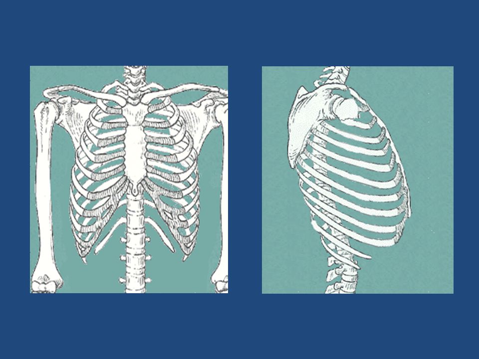
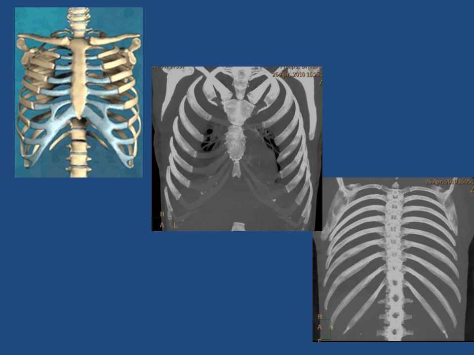
Toraks Duvarı Toraks duvarı hava geçirmeyen, genişleyebilen, koni şeklinde bir kafesdir. Toraks duvarının ön duvarını sternum; yanlarını ilk 10 kot kaplar. Arka göğüs duvarı, 12 torasik vertebra, bunların transvers çıkıntıları ve 12 kostadan oluşmaktadır. Torasik kafesin üst ön kısmı klavikula ve subklavian damarlar, lateralde omuz ve aksilla, sinir ve damarlar; dorsalde skapula tarafından çevrilir.

5
Kemik yapılar Kostalar Vertebralar Sternum Klavikula Skapula

6
Kostalar 12 çift kosta var.
İlk 7 kosta arkada vertebralarla, önde de kostosternal kıkırdak yapılarla sternumla birleşir. Bu kostalara gerçek ya da vertebro-sternal kosta denir Sonraki 3 kosta önde diğer kostaların ön bölümlerine yapışır: vertebro-kondral kosta Son iki kosta boştadır ve vertebral kosta denir

8
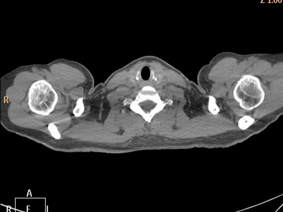
Kostalar -3 segmentten oluşur: baş,boyun ve gövde bölümü. Kosta tüberkülü gövdeden orijin alır. -Alt iç yüzde ‘kostal oluk’ var ve buradan sinir ve damarlar geçer. The tubercle of a rib is a projection located posteroinferior and lateral to the neck of the rib. It articulates with the transverse process of the vertebra of the same number. So, the tubercle of rib 7 should articulate with the transverse process of the T7 vertebra. The head of the rib is the part of the rib that articulates with the demifacets of two adjacent vertebral bodies. So, the head of rib 7 should articulate with the 6th vertebra superiorly and the 7th vertebra inferiorly.

9
Kostalar -Vertebra ile 2 sinoviyal eklemi var: Kostovertebral eklem (baş ile vertebra korpusu aynı ve bir üst seviye) Kostotransvers eklem (tüberkül ile aynı seviye vertebra korpusu) The tubercle of a rib is a projection located posteroinferior and lateral to the neck of the rib. It articulates with the transverse process of the vertebra of the same number. So, the tubercle of rib 7 should articulate with the transverse process of the T7 vertebra. The head of the rib is the part of the rib that articulates with the demifacets of two adjacent vertebral bodies. So, the head of rib 7 should articulate with the 6th vertebra superiorly and the 7th vertebra inferiorly.
 The tubercle of a rib is a projection located posteroinferior and lateral to the neck of the rib. It articulates with the transverse process of the vertebra of the same number. So, the tubercle of rib 7 should articulate with the transverse process of the T7 vertebra. The head of the rib is the part of the rib that articulates with the demifacets of two adjacent vertebral bodies. So, the head of rib 7 should articulate with the 6th vertebra superiorly and the 7th vertebra inferiorly.")
10
Kostotransvers eklem Kostovertebral eklem

11
Kostalar Nörovasküler demet:
-Ven, arter ve sinir kostanın alt ve iç kenarı boyunca seyreder. -Eğer girişim yapılacaksa kostanın üst kenarı tercih edilmelidir.

12
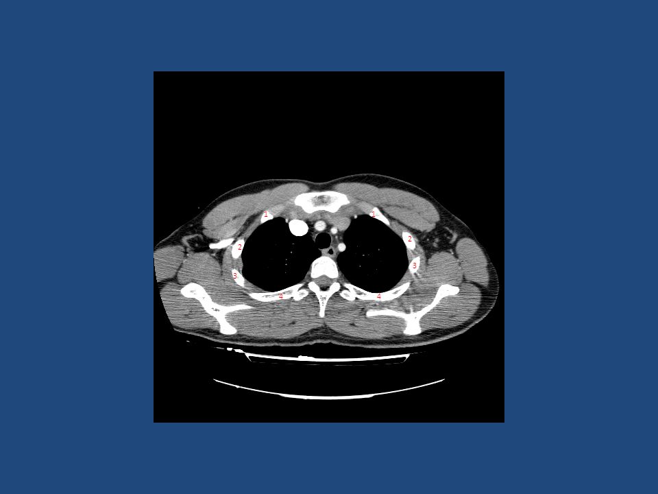
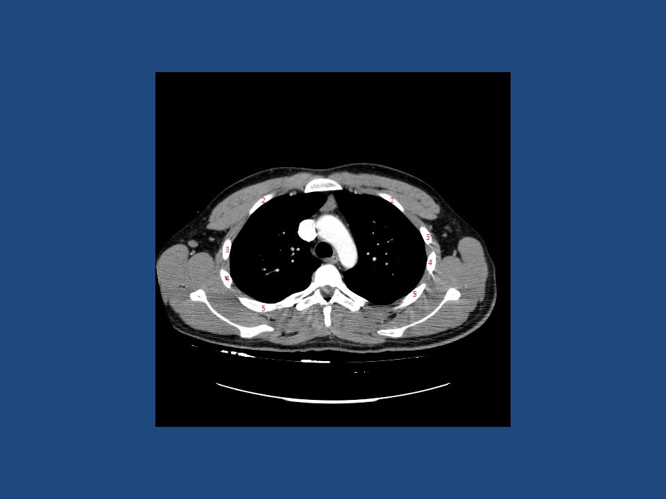
Kostalar Nörovasküler demet: -Her interkostal boşlukta 2 adet anterior interkostal arter bir adet posterior interkostal arter seyreder. -İlk 5-6 anterior interkostal arter internal torasik arter dallarından sonraki alt alanlar ise muskulofrenik arter dallarından beslenir -Posterior interkostal arterlerin ilk ikisi süperior interkostal arter dallarından sonrakiler inen aortadan dallanır.

13
Th1 1 1

14
1 1

15
1 1 2 2
16
2 2

17
2 2

18
2 2

19
2 2

20
2 2

21
2 2

22
2 2

23
2 2

24
2 2

25
2 2

26
2 2

27
2 2

28
2 2

29
2 2

30
2 2

31
2 2

32
1 1 2 2 3 3 4 4
33
2 2

34
2 2

35
2 2

36
2 2

37
2 2

38
2 2 3 3 4 4 5 5
40
Vertebralar Kostalara eşlik eden 12 vertebra vardır.
İçinden spinal kord geçer. Gövde, transver çıkıntı ve arka elemanlardan oluşur. Torasik 1. vertebra spinöz çıkıntısı dıştan palpe edilebilen en çıkıntılı ikinci vertebra (en çıkıntılısı C7 vertebra spinöz çıkıntısı)
")
42
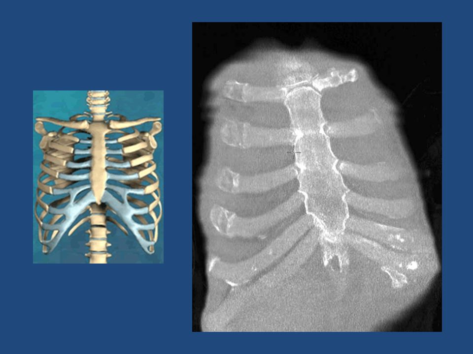
Sternum -Manubrium, gövde ve ksifoid bölümlerinden oluşur. -Sternal açı: İkinci kostal kartilajın sternuma yapıştığı nokta. Kalp oskültasyonu için kullanılır. Sternal çentik /juguar çentik is manubriumun klavikula ile eklem yaptığı üst kenar

44
Manubrium Manubriosternal eklem Gövde Ksifoid Kostal kartilaj

45
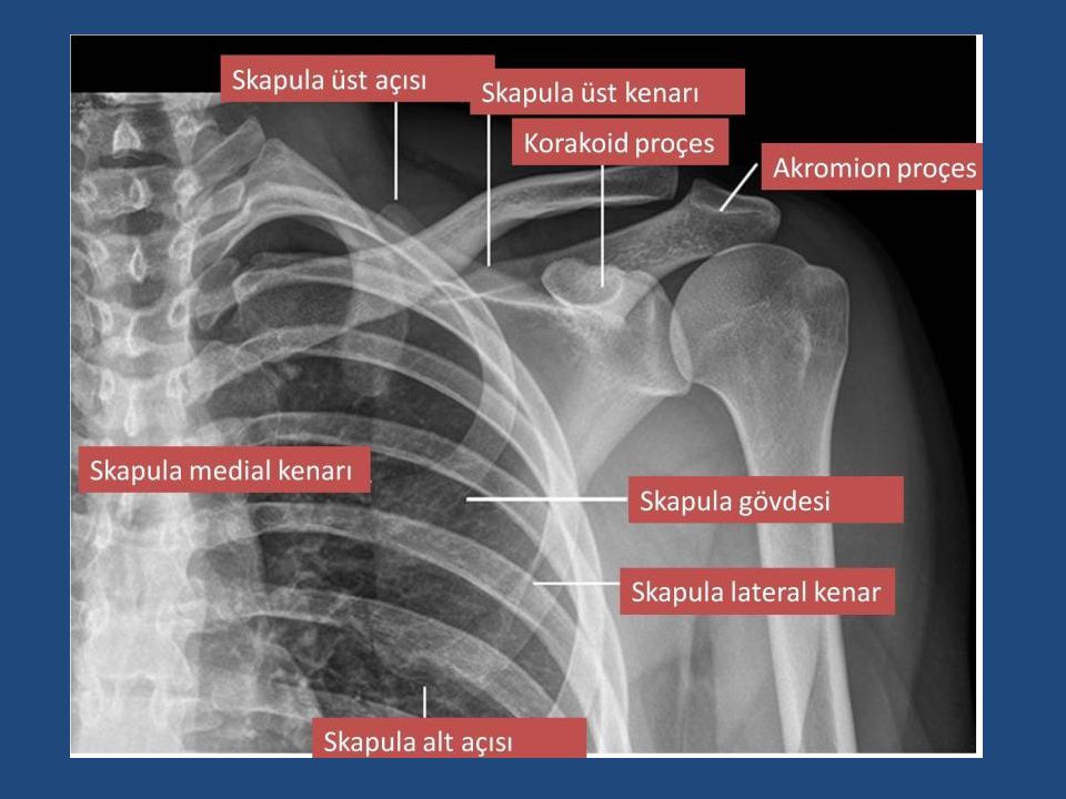
Skapula Gövde iskeletine yalnız kas ve bağlar yardımıyla tutunmuş üçgen bir kemiktir. Arka yüzünün 1/3 üst bölümünde “Spina skapula” denilen ve dış yana uzayan bir kemik çıkıntısı vardır. Bu kemik çıkıntının ucuna “Akromion” denir. Akromion klavikula kemiğinin dış ucuyla eklemleşir. Üst dış köşesinde humerus başıyla eklemleşen bir eklem yüzeyi vardır. Bu eklem yüzeyine “glenoid” denir. Üst kenarı, dış yanda “Korakoid çıkıntı” denilen bir çıkıntıyla sonlanır.

48
Klavikula

49
Sternoklavikuler eklem

50
Yumuşak dokular Süperior sulkus Kaslar Meme Cilt ve ciltaltı

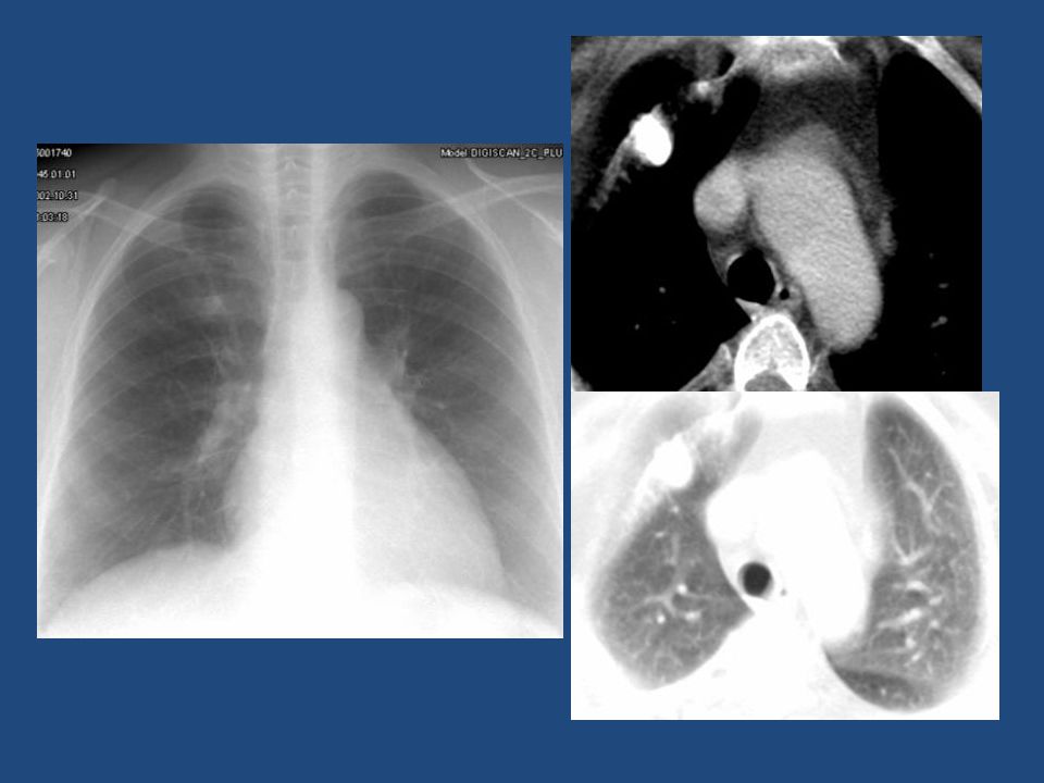
51
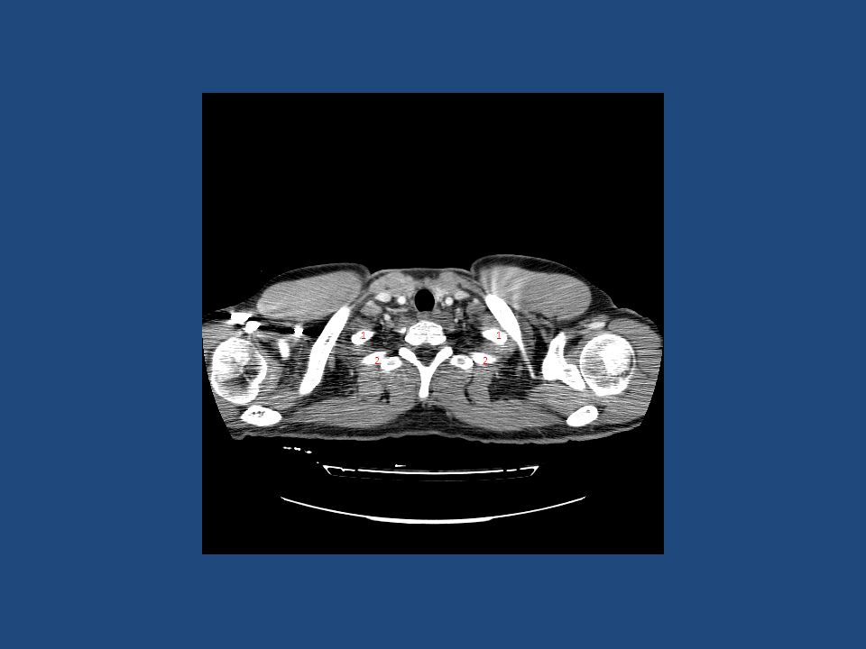
Süperior sulkus Akciğer apikal göğüs duvarında yeralan içinde subklaviyen damarlar, stellat ganglion ve brakial pleksusun sinir köklerinin geçtiği alan 3 skalen kas ön, orta ve arka olmak üzere 3 kompartmana ayırır. arrow indicates the stellate ganglion. L = lower trunk of brachial plexus, M = middle trunk of brachial plexus, SA = subclavian artery, SV = subclavian vein, U = upper trunk of brachial plexus.

52
Süperior sulkus Anterior kompartmanda SUBKLAVİYEN VEN
Orta kompartmanda SUBKLAVİYEN ARTER ve BRAKİAL PLEKSUS DALLARI Arka kompartmanda da STELLAT GANGLİON ve BRAKİAL PLEKSUS kökleri yeralır arrow indicates the stellate ganglion. L = lower trunk of brachial plexus, M = middle trunk of brachial plexus, SA = subclavian artery, SV = subclavian vein, U = upper trunk of brachial plexus.

53
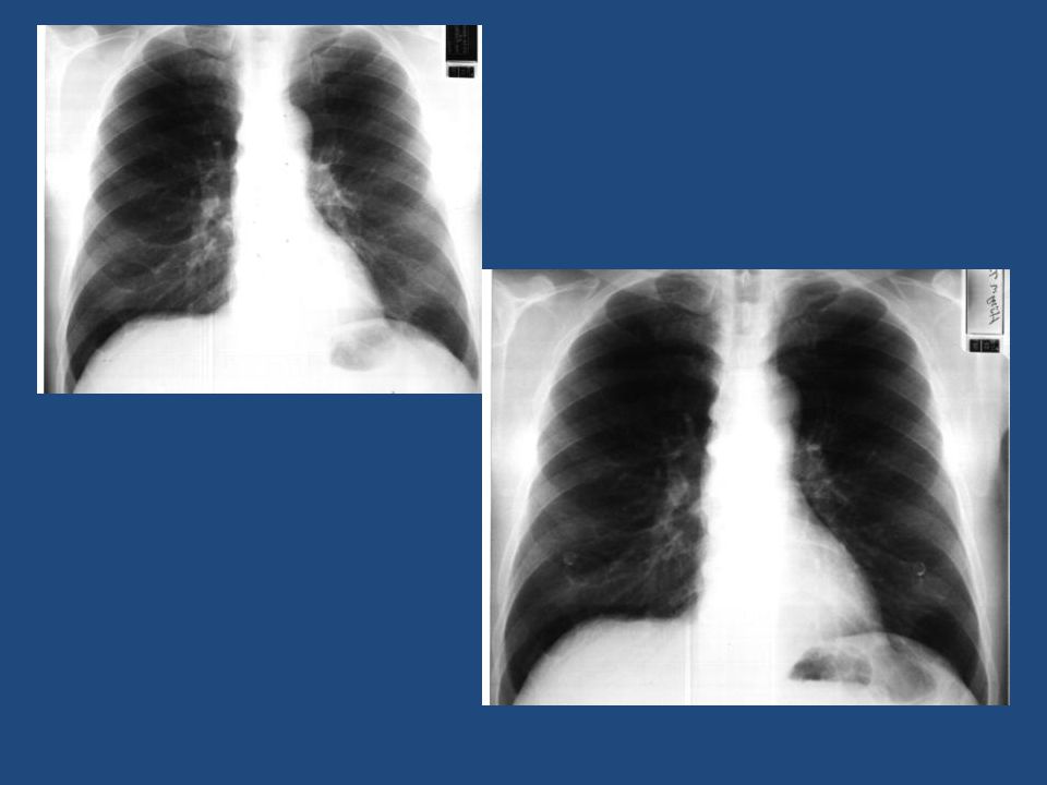
HER APİKAL TÜMÖR SÜPERİOR SULKUS (PANCOAST) TÜMÖR DEĞİLDİR
 TÜMÖR DEĞİLDİR")
54
Yumuşak dokular Parietal plevradan itibaren dışarı doğru katmanlar:
- Endotorasik (subplevral) fasya - Derin (innermost) interkostal kas - İnternal interkostal kas - Eksternal interkostal kas
 fasya. - Derin (innermost) interkostal kas. - İnternal interkostal kas. - Eksternal interkostal kas.")
55
Endotorasik fasya Toraks duvarını örten ekstraplevral seröz membran
Parietal plevrayı göğüs duvarı ve diyafragmadan ayırır Visseral plevra Parietal plevra Endotorasik fasya

56
İnterkostal kaslar - Derin (innermost) interkostal kas - İnternal interkostal kas - Eksternal interkostal kas
 interkostal kas - İnternal interkostal kas - Eksternal interkostal kas")
57
The muscles of the thorax consist of the intercostals and diaphragm
The muscles of the thorax consist of the intercostals and diaphragm. The intercostal muscles are arranged as three layers (external layer, internal layer and an incomplete innermost layer) between the ribs. The diaphragm closes the thoracic outlet and separates the thoracic cavity from the abdominal cavity. The three layers of the intercostal muscles are: external layer -- external intercostal internal layer -- internal intercostal innermost layer -- transversus thoracic (anterior), innermost (lateral) and subcostal (posterior) The diaphragm is the most important muscle of the thoracic wall. During normal respiration, this muscle is the primary component. As you can see, the innermost layer is split into three differently named muscle groups. The transversus thoracis, innermost intercostal and subcostal muscles make up the deepest layer of muscles from anterior to posterior, respectively.
 between the ribs. The diaphragm closes the thoracic outlet and separates the thoracic cavity from the abdominal cavity. The three layers of the intercostal muscles are: external layer -- external intercostal. internal layer -- internal intercostal. innermost layer -- transversus thoracic (anterior), innermost (lateral) and subcostal (posterior) The diaphragm is the most important muscle of the thoracic wall. During normal respiration, this muscle is the primary component. As you can see, the innermost layer is split into three differently named muscle groups. The transversus thoracis, innermost intercostal and subcostal muscles make up the deepest layer of muscles from anterior to posterior, respectively.")
58
BT kesitlerde, kostanın iç kenarında visseral plevra, parietal plevra, normal plevral sıvı ve
endotorasik fasya ve en içte derin interkostal kas kalınlığı 2 mmden incedir

59
Toraks duvarı kasları Bu kaslar hem inspiryuma hem de ekspiryuma katkısı olan kaslardır. Omuz , kol ve boyun ve batın ile de ilişkilidirler.

60
Toraks duvarı kasları İnspirasyon kasları ekspirasyon kasları
Diyafragma (%70) m.intercostalis interni m.intercostalis externi m.rectus abdominis internus m.pectoralis major m.quadratus lumbarum m.pectoralis minör m.transversus abdominis m.sternocleidomastoidus m.obliquus abdominus m.scalanius anterior m.serratus posterior inferior m.serratus anterior m.latissumus dorsi
 m.intercostalis interni. m.intercostalis externi m.rectus abdominis internus. m.pectoralis major m.quadratus lumbarum. m.pectoralis minör m.transversus abdominis. m.sternocleidomastoidus m.obliquus abdominus. m.scalanius anterior m.serratus posterior inferior. m.serratus anterior. m.latissumus dorsi.")
61
Pektoralis major kası Pektoralis minör kası Subskapularis kası Latissimus dorsi İnfraspinatus kası Romboid kas Trapezius kası

62
The serratus anterior is a muscle that originates on the surface of the upper eight ribs at the side of the chest and inserts along the entire anterior length of the medial border of the scapula. The serratus anterior is occasionally called the "boxer's muscle" because it is largely responsible for the protraction of the scapula—that is, the pulling of the scapula forward and around the rib cage that occurs when someone throws a punch. The serratus anterior also helps to stabilize the scapula. In addition, it assists in rotating the scapula (glenoid fossa) upward.
 upward..")
67
VARYASYONLAR

70
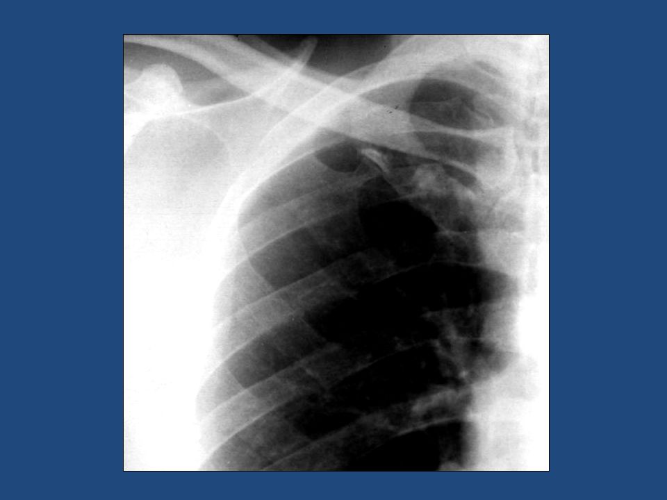
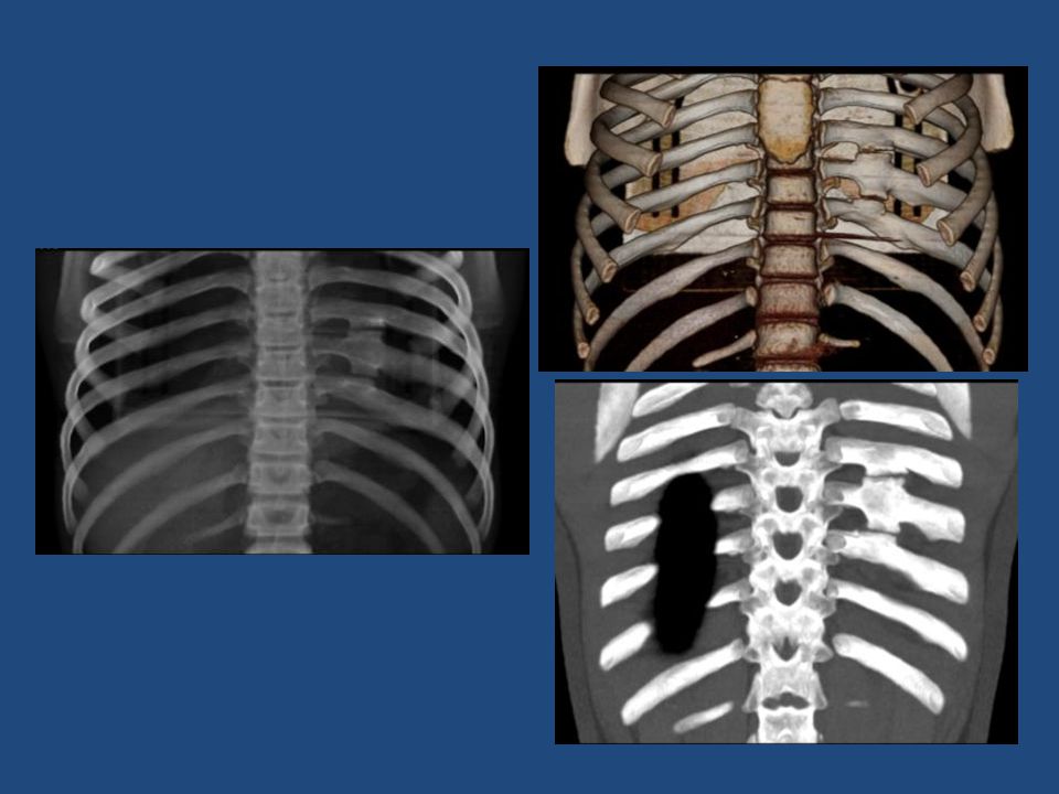
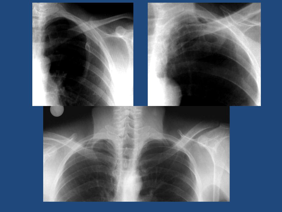
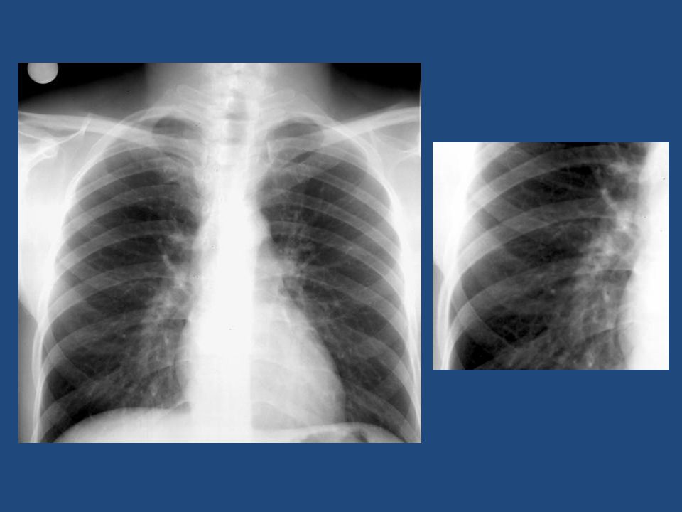
Romboid fossa: Kostoklavikuler ligaman
RHOMBOİD FOSSA: Klavikula ile 1. kot arasında yeralan kostoklavikuler ligamanın kuvveti ile ilişkili litik bir görünüm meydana gelir

71
Rhomboid fossa: The enlarged area (the thoracic inlet) shows defects in the under surface of the medial ends of the clavicles. Here it is more marked on the right, but they may be unilateral or bilaterally symmetrical also. They have been mistaken for lytic deposits, but actually represent a normal variant at the site of insertion of the rhomboid ligament between the first rib and the clavicle.

79
Episternal kemik

80
Midline sternal foramina

Benzer bir sunumlar