Sunuyu indir
Sunum yükleniyor. Lütfen bekleyiniz

1
Cekirdegin Yapisi

3
Fig. x-x Electron micrograph of nucleus
Fig. x-x Electron micrograph of nucleus. Note absence of condensed (hetero)chromatin under the pore. Also note the double membrane.
chromatin under the pore. Also note the double membrane.")
6
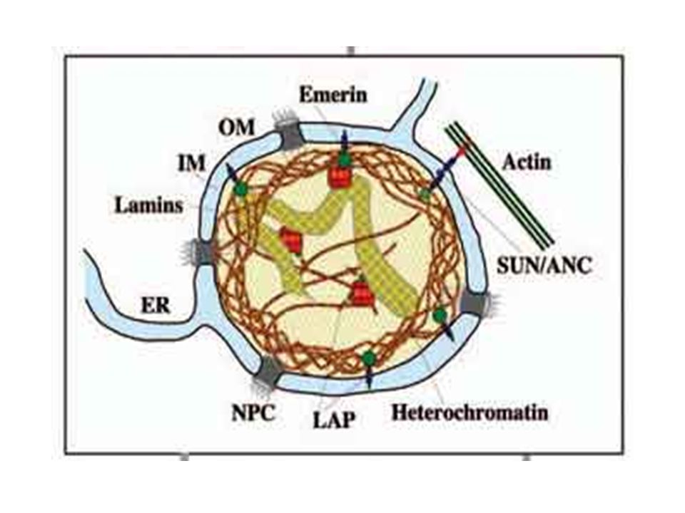
Nukleer kilifin ic membraninda katman boyunca agsi bir yapi olusur.
Nukleer lamina, nm. kalinliginda stabilize edici ara filamentlerle cevrili. Bu ara filamentler agirligi kD arasinda degisen lamin polimerleridir. Laminler cekirdegin fonksiyonel organizasyonunda gorevlidirler. Bunlar mitozdan once ve sonra katlanma ve acilmada gorevlidirler.

7
Laminler fosforile oldugu zaman, ayrisirlar
Laminler fosforile oldugu zaman, ayrisirlar. Nukleer kilif vezikullerine ayrilir. Defosforilasyon ise bu durumu tersine cevirir ve cekirdegin tekrar duzenlenmesi saglanir.

8
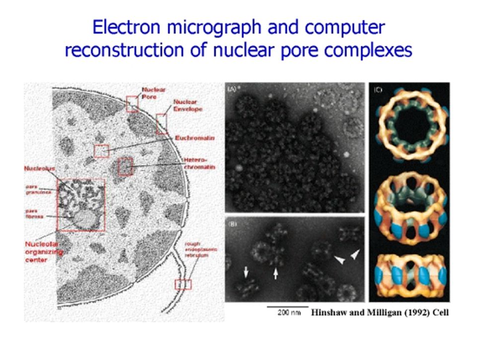
Nukleer Por Komplekslerinin SEM ile goruntulenmesi

9
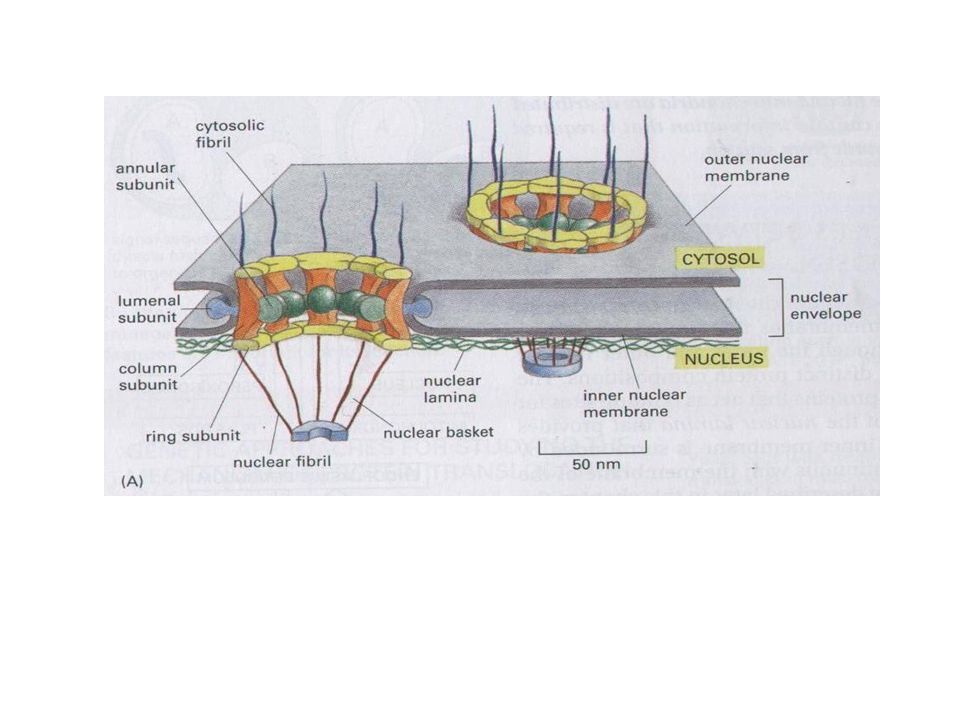
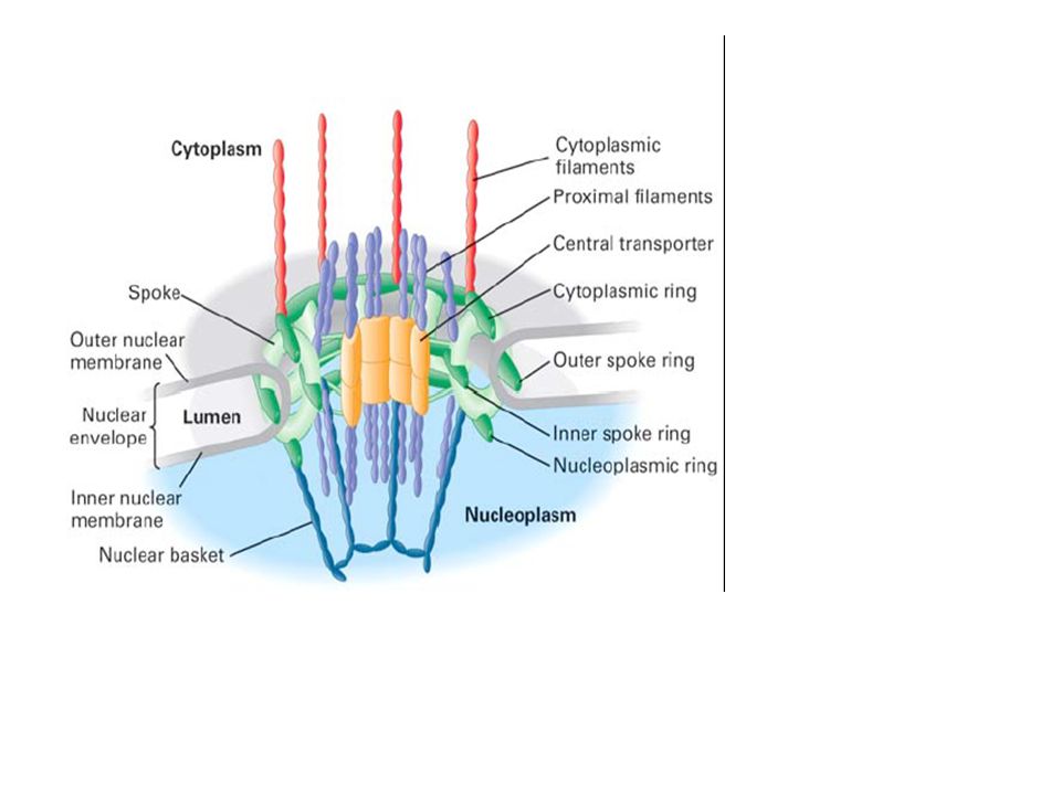
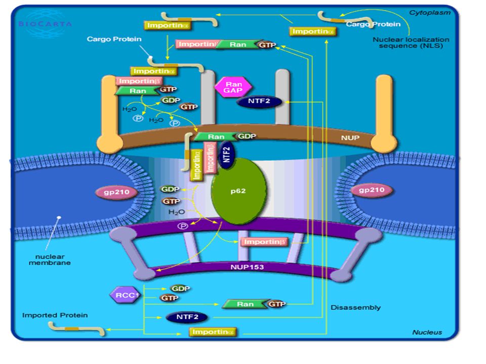
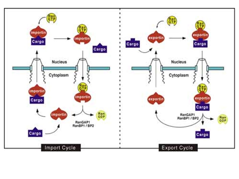
Nukleer Por Kompleksi

11
Nuclear pore coplex (by negative staining and resulting model).
.")
13
Nukleer por kompleksi mRNA’yi tanir ve secici olarak olgun mRNA’nin gecisine izin verir.
Nukleer kisitlayici proteinler (RNA poly II, snRNP’ler, hnRNP/ intron kompleksleri) nukleer cikistan hemen once uzaklastirilirlar. Mekik dokuyan proteinler(shuttling) mRNA’nin cikisina yardimci olmak icin bagli kalirlar.(hnRNP A1, SR proteinleri, CBC) mRNA molekulunun cekirdekten cikisi Ran-Bagimli degildir. Fakat tRNA ve snRNA’nin cikisi Ran-bagimlidir.
 nukleer cikistan hemen once uzaklastirilirlar. Mekik dokuyan proteinler(shuttling) mRNA’nin cikisina yardimci olmak icin bagli kalirlar.(hnRNP A1, SR proteinleri, CBC) mRNA molekulunun cekirdekten cikisi Ran-Bagimli degildir. Fakat tRNA ve snRNA’nin cikisi Ran-bagimlidir.")
15
Transport of mRNA out of the nucleus
Transport of mRNA out of the nucleus. Different proteins can be found associated with this abundant insect salivary gland mRNA inside and outside of the nucleus.

17
Fig 4-23 EM of nucleosomes (B) and the 30nm chromatin fibre of packed nucleosomes (A) in the normal conformation, which includes histone H1.
 and the 30nm chromatin fibre of packed nucleosomes (A) in the normal conformation, which includes histone H1.")
18
Nucleosome structure, except histone H1 which causes folding into the 30nm fibre.

19
Fig. x-x MODEL of DNA organization around nucleosomes.
Each human cell nucleus contains about 2m of DNA in 46 chromosomes packed into about 10um diameter nucleus.

20
Fig. 4-15 The size relationship between small human chromosome 22 (1
Fig The size relationship between small human chromosome 22 (1.5% of genome) and the gene number and size. There is a lot of repetitive and unused DNA.
 and the gene number and size. There is a lot of repetitive and unused DNA.")
21
STRUCTURE OF CHROMOSOMES AND DNA ORGANIZATION
Compare the highly condensed mitotic chromosome with relatively “loose” chromatin of interphase nucleus. But even interphase chromatin is HIGHLY ORGANIZED: supecoiled DNA and association with histone proteins in “nucleosomes”. In Eukaryotes, this is in a number of linear chromosomes, with general structure shown below:

22
Packaging DNA 11 nm 30 nm 200 nm 2 nm 700 nm Metaphase Chromosome
Looped Domains Nucleosomes B DNA Helix Tight helical fiber 700 nm G C A T Metaphase Chromosome Protein scaffold

Benzer bir sunumlar


![NATURAL BALANCE [Tabi Denge] Interest free system Destiny Social justice Balance in economics Balance in governance Balance in ethics Balance in knowledge.](/8/2539051/big_thumb.jpg "NATURAL BALANCE [Tabi Denge] Interest free system Destiny Social justice Balance in economics Balance in governance Balance in ethics Balance in knowledge.>")


>")


