Sunuyu indir
Sunum yükleniyor. Lütfen bekleyiniz

1
4 Boyutlu Ultrasonografi
Serdar H. Ural, M.D., F.A.C.O.G. Director, Division of Maternal Fetal Medicine Director, Labor and Delivery Unit Director, Fellowship Training Program Director, Obstetrical Ultrasound Suite Professor of Obstetrics & Gynecology and Radiology Penn State University College of Medicine, U.S.A.

2
3D/4D Ultrason 2D = 2 boyutlu 3D = 3 boyutlu
4D= 3 boyutlu canli (zamanli) real-time goruntu
 real-time goruntu.")
3
4D Ultrason 2D ultrason gebelik tetkikinde kullanilan belki de en onemli metottur 3D/4D ultrasonun yayginlasmasi, gebelik takibinde gerekli olup olmadiginin tartismasini yaratmistir 3D/4D ultrason, bilhassa 2D ile yapilan tetkikte tespit edilmis bazi anomalilerin daha iyi analiz edilmesinde faydali oldugu gorulmustur Fetal vucutta protruzyon yapan anomalilerin tetkikinde onemlidir- noral tup defekti, omfalosel, gastroskisis Cardiac American Institute of Ultrasound in medicine. Acoustic output measurement standards for diagnostic ultrasound equipment. Laurel (MD): AIUM; 1998 .
: AIUM;")
4
4D Ultrason Ultrasonun guvenilirligi uzun zamandan beri bilinmektedir
Insan calismalarinda yan etki gorulmemistir Bazi hayvan deneylerinde yani etki gorulmusse de bu calismalar baskalari tarafindan tekrarlandiginda ayni sonuclar elde edilmemistir Bilhassa 3D/4D ultrasonda termal endeks ve de mekanik endeks otomatik olarak kontrol edilmekte, ultrason muayenesi sirasinda dokuya yayilan enerji miktari minimumda tutulmaktadir Stark CR et al. Short and long term risks after exposure to diagnostic ultrasound in utero. Obstet Gynecol 1984; 63;

5
3D/4D Ultrason 3D popularitesi artmakta
iU22 (Phillips), Prosound Alfa-10 (Aloka), Voluson 730 Expert (GE) Ortalama muayene suresi 21 dakika 2D mean termal indeks (TI) , mekanik indeks (MI) – 1.12 3D mean TI , MI – 0.89 4D mean TI 3D/4D muayenesi 2.5 dakika ek Akustik seviye, enerji seviyesi yaklasik ayni Sheiner E et al. A comparison between acoustic output indices in 2D and 3D/4D ultrasound in obstetrics. Ultrasound Obstet Gynecol 2007, 29 (3); 326-8
, Prosound Alfa-10 (Aloka), Voluson 730 Expert (GE) Ortalama muayene suresi 21 dakika. 2D mean termal indeks (TI) , mekanik indeks (MI) – D mean TI , MI – D mean TI D/4D muayenesi 2.5 dakika ek. Akustik seviye, enerji seviyesi yaklasik ayni. Sheiner E et al. A comparison between acoustic output indices in 2D and 3D/4D ultrasound in obstetrics. Ultrasound Obstet Gynecol 2007, 29 (3);")
6
2D/3D/4D Ultrason 2D ultrason ve fetal fizyolojik cevap 100 hasta
18-24 gebelik haftasi, Fetal kalp atis hizi AF dikey olcumu Umbilical arter Doppler Muayene suresi Amniyotik sivi da azalma- Istatistiki fark (+) Fetal kalp hizinda artis- Istatistiki fark (-) Doppler, sure- Istatistiki fark (-) Akut fetal hemodinamik degisiklikler Ural SH, Repke J et al. Does 2nd trimester ultrasound effect normal fetal physiology. Oral Presentation, International Society of Ultrasound in Obstetrics and Gynecology Chicago, USA, Agustos 2008
 Fetal kalp hizinda artis- Istatistiki fark (-) Doppler, sure- Istatistiki fark (-) Akut fetal hemodinamik degisiklikler. Ural SH, Repke J et al. Does 2nd trimester ultrasound effect normal fetal physiology. Oral Presentation, International Society of Ultrasound in Obstetrics and Gynecology. Chicago, USA, Agustos")
7
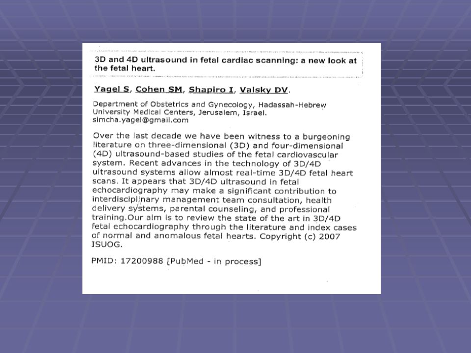
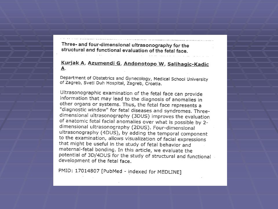
How Useful Is 3D and 4D Ultrasound in Perinatal Medicine
3D/4D hakkinda tip literaturunde su ana kadar yayinlanmis 575+ makale/calisma var, 438+ tanesi sirf 3D/4D hakkinda Yuz anomalileri Noral tup defektleri Iskelet anomalileri Konjenital kalp defektleri Intrakraniyel anomaliler Davranis Kurjak A et al. How useful is #d and 4D ultrasound in perinatal medicine. J Perinat Med 2007, 35 (1); 10-27 Martin J et al. Births; final data for Natl Vital Stat rep 2003, 52 (10):1-113
; Martin J et al. Births; final data for Natl Vital Stat rep 2003, 52 (10):")
8
3D/4D Ultrason 3472 fetal anatomik tarama, 2D ve 3D
906 tanesinde 1-5 anomali 2D ile karsilastirildiginda 3D - multiplanar tomografik inceleme %70 daha avantajli Defektin ciddiyetinin ve de normalitenin degerlendirilmesinde 2D’den daha mi iyi Long G et al. A comparative study of routine vs. selective fetal anomaly scanning. J Med Screen 1998; 5; 6-10

9
3D/4D Ultrason 99 fetus, once 3D/4D, sonrasinda 2D
2D 54 fetus normal, 45 fetus’ta 82 anomali 2D ve 3D/4D arasinda anomali tanisinda anlasma orani %90, intraclass correlation coefficient, 0.834; %95 CI, 3D/4D 2D’den 6 eksik anomali teshis etmis VSD (2) IVC blokaji Tetralogy of Fallot Bobrek Cystic adenmatoid malformasyon Dogum sonrasi tani ile karsilastirma, sensitivite/spesifisite 2D %96 - %73 3D/4D %92 - %76 Istatistiki olarak ciddi fark yok Goncalves L et al. What does 2 dimensional imaging add to 3D/4D obstetric ultrsound. J Ultrasound Med 2006, 25 (6); 691-9 Long G et al. A comparative study of routine vs. selecyive fetal anomaly scanning. J Med Screen 1998; 5; 6-10
 IVC blokaji. Tetralogy of Fallot. Bobrek. Cystic adenmatoid malformasyon. Dogum sonrasi tani ile karsilastirma, sensitivite/spesifisite. 2D %96 - %73. 3D/4D %92 - %76. Istatistiki olarak ciddi fark yok. Goncalves L et al. What does 2 dimensional imaging add to 3D/4D obstetric ultrsound. J Ultrasound Med 2006, 25 (6); Long G et al. A comparative study of routine vs. selecyive fetal anomaly scanning. J Med Screen 1998; 5;")
11
3D/4D Ultrason 13-34 gebelik haftalarinda
3D/4D ile kalp fonksyon tesbiti Tomografik real-time 3D Sistolik/diyastolik, atriyal/ventrikuler hemodinamik tesbit Anatomik anomali gorulmese de kalp anomalilerine bagli degisiklikler Hata T et al. 4D ultrasound in spatiotemporal image correlation for fetal heart visualization. J Clin Ultrasound Mayis 2008;36;4;204-7.

14
3D/4D Ultrason The effects on maternal anxiety of 2D vs. plus 3D/4D ultrasound in pregnancies at risk of fetal abnormalities; A randomized study 4D ultrason uygulanan annelerin %80’i, 2D ile karsilastirildiginda, bebegin normal oldugunun daha inandirici oldugunu Fakat anksiyetenin istatistiki olarak daha dusuk olmadigi

15
3D/4D Ultrason 2D ve 3D/4D ultrason kullanildiginda annenin bebegine psikolojik bagi Yayinlanmis bazi calismalarda bu bagin ultrasonla guclendigi, ve de uzun vadede anne ve bebegin daha az hastaliga yakalanmasina sebep olabilecegi varsayilmistir 3D/4D ultrason daha mi etkili Penn State Universitesinde calisma Rados C. FDA cautions against ultrasound keepsake images. FDA Consum 2004; 38 (1); 12-6
;")
16
Ultrasound Research Symposium
Nisan, 2010 Philadelphia Best Research Prize

17
Does Ultrasound Influence the Bond between Mother and Fetus?
Serdar Ural, MD, and MFM Team Penn State University, College of Medicine Introduction Maternal-fetal attachment (MFA) is a new concept that encompasses the behaviors mothers evoke toward their babies. A handful of studies have been conducted to determine if fetal ultrasounds are a means for increasing or even inducing a mother’s attachment towards her unborn child. Many studies have compared the effects of 3D and 4D ultrasounds on MFA, however, few have investigated the influence of the basic 2D ultrasound, considering it is the most common type of scan used in practice. It is hypothesized that the ultrasound experience will have a positive impact on a mother’s feelings for her fetus by increasing her attachment to the fetus. Methods 100 women obtaining a routine ultrasound scan at the Department of Maternal-Fetal Medicine at Hershey Medical Center were recruited. The 25-question survey given before and after the ultrasound was based on the MFA questionnaire developed by Cranley with additional statements addressing prenatal care. Group A (64 patients) completed a 21-question survey. Group B (35 patients) completed a 25-question survey, which included 4 questions on prenatal care. The difference between the pre-ultrasound mean and post-ultrasound mean was analyzed by a nonparametric 1-sample Wilcoxon signed ranks test. Figures Results The data from the entire study population (Group A, 100 patients) was analyzed based on 21 questions while the data of 35 patients (Group B) was analyzed based on 25 questions. Group A and Group B both resulted in a p value <.0001 indicating that the difference in MFA scores before and after the ultrasound is statistically significant. Questions 22-25, assessing prenatal care, were analyzed individually. Question 23 was statistically significant with a p value of Conclusions The ultrasound experience is one of many major events that occur during pregnancy. It becomes a lasting memory, especially if it is the first pregnancy. This study determined that 2D ultrasound significantly increases the bond between mother and fetus in addition to 3D and 4D imaging. The fact that ultrasound has such a profound impact on the thoughts of mothers suggests that it can be a stepping stone to incorporate other aspects of prenatal care into the appointment. Confounding variables: multiple ultrasound technicians and less than ideal results of anatomical survey. Future research: father’s reaction to ultrasound experience, the effect of other patients having an ultrasound on the MFA, the use of ultrasound to prevent maternal use of alcohol and tobacco. Table 1: Analysis of Group A maternal-fetal attachment scores (Q1-Q21) Observations 100 Mean 0.130 Std Deviation 0.182 Wilcoxon Signed Rank Statistic 1469.5 p value <.0001 Table 2: Analysis of Group B maternal-fetal attachment scores (Q1-Q25) Observations 35 Mean 0.181 Std Deviation 0.208 Wilcoxon Signed Rank Statistic 198 p value <.0001 Table 3: Analysis of prenatal care questions 22-25 Wilcoxon Statistic p value Question 22 3 0.25 Question 23 14 0.0156 Question 24 1.5 0.5 Question 25 -1 1.0
 is a new concept that encompasses the behaviors mothers evoke toward their babies. A handful of studies have been conducted to determine if fetal ultrasounds are a means for increasing or even inducing a mother’s attachment towards her unborn child. Many studies have compared the effects of 3D and 4D ultrasounds on MFA, however, few have investigated the influence of the basic 2D ultrasound, considering it is the most common type of scan used in practice. It is hypothesized that the ultrasound experience will have a positive impact on a mother’s feelings for her fetus by increasing her attachment to the fetus. Methods. 100 women obtaining a routine ultrasound scan at the Department of Maternal-Fetal Medicine at Hershey Medical Center were recruited. The 25-question survey given before and after the ultrasound was based on the MFA questionnaire developed by Cranley with additional statements addressing prenatal care. Group A (64 patients) completed a 21-question survey. Group B (35 patients) completed a 25-question survey, which included 4 questions on prenatal care. The difference between the pre-ultrasound mean and post-ultrasound mean was analyzed by a nonparametric 1-sample Wilcoxon signed ranks test. Figures. Results. The data from the entire study population (Group A, 100 patients) was analyzed based on 21 questions while the data of 35 patients (Group B) was analyzed based on 25 questions. Group A and Group B both resulted in a p value <.0001 indicating that the difference in MFA scores before and after the ultrasound is statistically significant. Questions 22-25, assessing prenatal care, were analyzed individually. Question 23 was statistically significant with a p value of Conclusions. The ultrasound experience is one of many major events that occur during pregnancy. It becomes a lasting memory, especially if it is the first pregnancy. This study determined that 2D ultrasound significantly increases the bond between mother and fetus in addition to 3D and 4D imaging. The fact that ultrasound has such a profound impact on the thoughts of mothers suggests that it can be a stepping stone to incorporate other aspects of prenatal care into the appointment. Confounding variables: multiple ultrasound technicians and less than ideal results of anatomical survey. Future research: father’s reaction to ultrasound experience, the effect of other patients having an ultrasound on the MFA, the use of ultrasound to prevent maternal use of alcohol and tobacco. Table 1: Analysis of Group A maternal-fetal attachment scores (Q1-Q21) Observations Mean Std Deviation Wilcoxon Signed Rank Statistic p value. < Table 2: Analysis of Group B maternal-fetal attachment scores (Q1-Q25) Observations. 35. Mean Std Deviation Wilcoxon Signed Rank Statistic p value. < Table 3: Analysis of prenatal care questions Wilcoxon Statistic. p value. Question Question Question Question")
18
4D Ultrason Prenatal invaziv tani ve tedavisi icin 4D, 93 fetus
Amniosentez, amnioinfuzyon, CVS, kordosentez Prosedur mean 5 dakika, %100 basari Oligohidramniyoz, ince plasenta, ince kord Zaman, komplikasyon riski daha dusuk Kim S et al. 4D ultrasound guidance of prenatal invasive procedures. Ultrasound Obstet Gynecol 2005, 26 (6); 663-5
;")
19
Maternal Obezite ve Fetal Anomali Taramasi 2D
Fetal anatomy ultrasound screening American Institute of Ultrasound in Medicine (AIUM) Over 25 structures 18-22 weeks of gestation Variable sensitivity 34-60% Decreases with increased BMI Absence of markers, 80% reduction in Down syndrome risk Experience Standardization
 Over 25 structures weeks of gestation. Variable sensitivity % Decreases with increased BMI. Absence of markers, 80% reduction in Down syndrome risk. Experience. Standardization.")
20
Obezite 2D Suboptimal visualization Significant ultrasound impairment
Visualization decreases Mostly cardiac and spine + others Suboptimal visualization Obezite 17%

21
Maternal Obezite ve Fetal Anomali Taramasi 3D/4D
18-24 hafta 11,000+ vaka Body mass index (BMI) >25 BMI >25, >30, >40 Sensitivite %66’dan %49, %25’lere inmis 3D/4D yarari? Dashe et al. Obstetrics/Gynecology, Mayis, 2009.
 >25. BMI >25, >30, >40. Sensitivite %66’dan %49, %25’lere inmis. 3D/4D yarari Dashe et al. Obstetrics/Gynecology, Mayis,")
22
3D/4D Ultrason 4D = 3 boyutlu canli (zamanli) real-time goruntu
4D = tomografik ultrason incelemesi Hacim olcumu icin ideal Operatif prosedurlerde daha guvenilir Hasta/fetus’e yan etki kaniti yok 3D/4D yayinlanmis calisma sayisi az 2D + 3D/4D su anda en faydali yontem Tek basina 3D/4D muayene icin yeterli veri yok

23
4D Ultrason & Davranis Bugune kadar yapilan deney ve calismalarda fetal davranis bicimlerinin ultrason ile incelenmesi sonucu fetal saglik hakkinda bilgi verip veremeyecegi yonundedir Bu calismalarda fetal davranis dendiginde el kol hareketlerinden tutun yuz ifade degisikligine kadar bircok hareket edebilen bolge ele alinmaktadir Fetal davranis incelenmesinin en iyi bilineni ve standart yontemi olan biyofizik profildir (BPP) Burda el, kol, govde gibi hareket edebilen bolgelerin incelenmesi sonucu evet fetus saglikli, ya da hayir fetus saglikli degil, dogurmak gerekir gibi sonuclara ulasmaktayiz
 Burda el, kol, govde gibi hareket edebilen bolgelerin incelenmesi sonucu evet fetus saglikli, ya da hayir fetus saglikli degil, dogurmak gerekir gibi sonuclara ulasmaktayiz.")
24
BPP Fetal hareket = 2 Fetal tonus = 2 Solunum = 2 Amniyotik sivi = 2
NST = 2 Toplam 10/10

25
4D Ultrason & Davranis 14 hareket 4D ile incelenebilir; Goz kapagi
Agiz Dil Dudak bukme Gulumseme Somurtma Esneme El’in bas’a El’in yuze (goz, kulak, agiz) Bas antefleksyon/retrofleksyon Emme Yutkunma
 Bas antefleksyon/retrofleksyon. Emme. Yutkunma.")
26
4D Ultrason & Davranis Neonatal donemdeki yuz ifadeleri incelendiginde fetal surecte 4D ile gorulenlerle ayni oldugu tespit edilmis Dogum sonrasi stres’te olan bebegin yuz ifadeleri, fetal donemde stres’te olan bebegin ifadeleriyle benzerlik gostermekte Fetal noral gelisim hakkinda bilgi vermekte Santral sinir sisteminin gelisimi direk olarak bu davranislara yansir Spesifik hareket duzeni (SMP) mevcuttur
 mevcuttur.")
27
4D Ultrason & Davranis Fetal davranisin dinamik analizi, santral sinir sisteminin maturasyonu ve gelismesi ile direk baglantilidir Norolojik hastaliklarin erken teshisi

28
4D Ultrason & Davranis Cok az data Arastirma ve dergi yayin kalitesi?
Standart olusturmak icin cok erken Tekrarlanabilir sonuclar gormek sart Ornegin BPP Yani konumuz cok yeni ve henuz gelismekte olup literaturde temsili az

29
4D Ultrason Yani fetal davranis sadece tibbi bilgi vermekle kalmayip psikolojik ve emosyonel yani da var

30
4D Ultrason & Davranis Intrauterin gelisme geriligi (IUGR) oldugu bilinen fetuslerde fetal yuz ifadesi ve de vucut hareket kalitesinde degisikler oldugu tespit edilmis Bu da acaba beyin gelisiminde aksakliktan oturu olabilecegi teorisini gelistirmis Yani IUGR olan fetuslerde oksijenlenme aksakligi beyni etkileyip indirek yolla fetal davranisi da etkileyebilmektemidir? Bu da bize davranissal tanida yardimci olabilirmi sorusunu sordurtmaktadir
 oldugu bilinen fetuslerde fetal yuz ifadesi ve de vucut hareket kalitesinde degisikler oldugu tespit edilmis. Bu da acaba beyin gelisiminde aksakliktan oturu olabilecegi teorisini gelistirmis. Yani IUGR olan fetuslerde oksijenlenme aksakligi beyni etkileyip indirek yolla fetal davranisi da etkileyebilmektemidir Bu da bize davranissal tanida yardimci olabilirmi sorusunu sordurtmaktadir.")
31
Fetal Behaviour of IUGR Fetuses by 4D USG
Fetal yuz ifadesi, vucut hareket kalitesi Beyin gelisiminde aksaklik? 50 IUGR, 50 kontrol Hareket duzeni tamamen farkli, hareketlerde farklilik IUGR – azalmis hareket, sayi, bicim, duzen El’in bas’a hareketi El’in yuz’e hareketi Bas retrofleksyon Andonotopo W et al. The assessment of fetal behaviour of growth restricted fetuses by 4D ultraound. J Perinatol Med 2006, 34 (6);471-8
;")
32
4D Ultrason & Davranis Anensefalik fetus’te motor hareketlerde anormallikler IUGR’da benzer hareket degisiklikleri

33
Fetal Behaviour of IUGR Fetuses by 4D
Dogum oncesi bilgi icin Iyi fizyolojik bilgi 4D 3D’nin canli gosterimidir, 2D’ye gore daha iyi? Standart henuz yok

34
4D Ultrason & Davranis Eger tedavisi olan bir risk ise bilhassa biyofizik profil ile davranissal degisiklikler takip edilerek fetus’te duzelme olup olmadigi gorulebilir Fakat bunun standart olarak kullanilmasi henuz erkendir Daha fazlaca sayida kaliteli yayin gerekmektedir Cok merkezli calismalara acilen ihtiyac vardir Bu sayede fetal norodavranis ve cocuk gelisimsel sonuclari normativ data elde edilerek, aralarindaki prediktebilite teyit edilmelidir

35
3D/4D Ultrason 4D = 3 boyutlu canli (zamanli) real-time goruntu
4D = tomografik ultrason incelemesi Hacim olcumu icin ideal Operatif prosedurlerde daha guvenilir Hasta/fetus’e yan etki kaniti yok 3D/4D yayinlanmis calisma sayisi az 2D + 3D/4D su anda en faydali yontem Tek basina 3D/4D muayene icin yeterli veri yok

36
Sonuc 2D + 3D/4D rutin kullanilmasi gebelikte en faydali yaklasim
Guvenilir, yan etki yok Anne-bebek bagi ve anne anksiyete 3D/4D belli anomalilerin incelenmesinde daha faydali Yumusak doku, protuzyon, kalp

37
Sonuc 4D’den elde edilen tomografik goruntuler bilgisayar araciligiyle anatomik reconstruction’dan gecip 2D ve 3D’ye gore organlar/olcumler daha net goruntu verecek 4D muayenesi hasta icin cok kisalacak Anomali ve de normal teshislerin dogruluk orani %100’e yaklasacak

38
Referans American Institute of Ultrasound in medicine. Acoustic output measurement standards for diagnostic ultrasound equipment. Laurel (MD): AIUM; 1998 Stark CR et al. Short and long term risks after exposure to diagnostic ultrasound in utero. Obstet Gynecol 1984; 63; Rados C. FDA cautions against ultrasound keepsake images. FDA Consum 2004; 38 (1); 12-6 Martin J et al. Births; final data for Natl Vital Stat rep 2003, 52 (10):1-113 Long G et al. A comparative study of routine vs. selecyive fetal anomaly scanning. J Med Screen 1998; 5; 6-10 Ewigman B et al. Effect of prenatal ultrasound screening on perinatal outcome. RADIUS study froup. N Engl J Med 1993; 329; 821-7 Kurjak A et al. How useful is #d and 4D ultrasound in perinatal medicine. J Perinat Med 2007, 35 (1); 10-27 Sheiner E et al. A comparison between acoustic output indices in 2D and 3D/4D ultrasound in obstetrics. Ultrasound Obstet Gynecol 2007, 29 (3); 326-8 Goncalves L et al. What does 2 dimensional imaging add to 3D/4D obstetric ultrsound. J Ultrasound Med 2006, 25 (6); 691-9 Kim S et al. 4D ultrasound guidance of prenatal invasive procedures. Ultrasound Obstet Gynecol 2005, 26 (6); 663-5 Leung et al. The effects on maternal anxiety of 2D vs. plus 3D/4D ultrasound in pregnancies at risk of fetal abnormalities; A randomized study. Ultrasound Obstet Gynecol 2006, 28 (3):
: AIUM; Stark CR et al. Short and long term risks after exposure to diagnostic ultrasound in utero. Obstet Gynecol 1984; 63; Rados C. FDA cautions against ultrasound keepsake images. FDA Consum 2004; 38 (1); Martin J et al. Births; final data for Natl Vital Stat rep 2003, 52 (10): Long G et al. A comparative study of routine vs. selecyive fetal anomaly scanning. J Med Screen 1998; 5; Ewigman B et al. Effect of prenatal ultrasound screening on perinatal outcome. RADIUS study froup. N Engl J Med 1993; 329; Kurjak A et al. How useful is #d and 4D ultrasound in perinatal medicine. J Perinat Med 2007, 35 (1); Sheiner E et al. A comparison between acoustic output indices in 2D and 3D/4D ultrasound in obstetrics. Ultrasound Obstet Gynecol 2007, 29 (3); Goncalves L et al. What does 2 dimensional imaging add to 3D/4D obstetric ultrsound. J Ultrasound Med 2006, 25 (6); Kim S et al. 4D ultrasound guidance of prenatal invasive procedures. Ultrasound Obstet Gynecol 2005, 26 (6); Leung et al. The effects on maternal anxiety of 2D vs. plus 3D/4D ultrasound in pregnancies at risk of fetal abnormalities; A randomized study. Ultrasound Obstet Gynecol 2006, 28 (3):")
Benzer bir sunumlar


>")
>")


