Sunuyu indir
Sunum yükleniyor. Lütfen bekleyiniz

1
CoolFit tanıtım sunumudur. ilerlemek için tıklayınız

4
CoolFit uygulama öncesi ve sonrası yağ hücreleri…
Uygulamadan 16 gün sonra This slide shows some of the early histology produced at Mass General during the early research on cryolipolysis on pigs. This view shows the untreated control portion. And this is a close-up of the fat immediately under the skin. Note the healthy adipocytes that make up the fat lobules (point to the healthy lobules). A few inches away, on the same pig, the tissue was treated. This slide shows the condition of the fat 16 days after treatment. As you can see, the adipocytes in these lobules (point to one of the lobules) have been killed, and the inflammatory cells have infiltrated the region. These cells are lymphocytes, histiocytes and macrophages. At this stage, some of the fat has been removed, but much remains to be carried away through the lymph system by the inflammatory process.
. A few inches away, on the same pig, the tissue was treated. This slide shows the condition of the fat 16 days after treatment. As you can see, the adipocytes in these lobules (point to one of the lobules) have been killed, and the inflammatory cells have infiltrated the region. These cells are lymphocytes, histiocytes and macrophages. At this stage, some of the fat has been removed, but much remains to be carried away through the lymph system by the inflammatory process.")
5
CoolFit uygulama sonrası yağ hücreleri…
Lipolysis Immediate 3 days 7 days Inflammation 14 days 30 days 60 days This is the most important science slide I have to show you. It describes what happens to the fat over time after a Zeltiq Procedure. First, let me describe the pig study that we did to get this histology series. This was a 90-day study. At the end of the study, the animal was sacrificed and all the tissue was harvested for pathology. 90 days prior to that, one small area was treated on the pig’s abdomen. (Gesture with your hands, pointing to a spot on your chest where the first treatment was.) 60 days prior to the end of the study, a second area was treated on the same animal adjacent to the first treatment. (Move your finger, pointing to a place on your chest 6 inches from the first treatment location.) Then, 30 days prior, 14 days, and so on. (Focus them back to the PowerPoint now. Point to the “immediate” histology.) This is the fat that was treated just before the animal was sacrificed. In each of these pictures, they represent the fat immediately under the dermis, and the dermis is off to the right side of the picture. As you can see from this histology, this fat looks COMPLETELY NORMAL. In fact there is nothing about this sample that looks any different from a control sample (which is not shown). This is because the cells have been injured in a subtle way that is not visible even with this type of histology. The cells are still alive, but they have been fatally injured. The procedure initiates the apoptotic cascade, and all the fat cells in this picture will die within the next 2 to 3 days. (Now, pointing to the 3-day histology…) By 3 days post-treatment, all these cells have died through apoptosis. The first histological evidence of that is now evident as inflammatory cells begin to infiltrate the fat. The tiny blue dots you see here are the nuclei of the inflammatory cells. (Pointing to the 7-day pic…) At 7 days post, the inflammation increases… And if I blow up this section you can see the dead fat cell… …And the nuclei of the inflammatory cells at higher magnification. The inflammation continues to intensify at 14 and 30 days. By 30 days (point to that pic) the fat cells are beginning to appear misshapen and the cells continue to be moved out of the subcutaneous tissue through the lymph system. By 90 days (point to the 90-day pic) most of the fat has been removed, but you can still see a little inflammation left, indicating that the process is not yet complete, and more fat will be removed. You will also note that there is a significant change in the density of the fibrous septae, the collagen, in the region immediately under the dermis. That is NOT because we have created scar tissue. All that collagen is the natural pre-existing collagen that was there before the treatment. But it has moved from deeper subcutaneous tissue as the adipocytes have been evacuated. (During that sentence, gesture with your hands showing that the fat came from about 6X below the field of view of that pic. If they ask how you know it isn’t new collagen, you can respond that we have stained for new collagen and found the treated areas have no more new collagen than the untreated control samples.) CoolFit uygulama sonrası yağ hücreleri… 90 days
 60 days prior to the end of the study, a second area was treated on the same animal adjacent to the first treatment. (Move your finger, pointing to a place on your chest 6 inches from the first treatment location.) Then, 30 days prior, 14 days, and so on. (Focus them back to the PowerPoint now. Point to the immediate histology.) This is the fat that was treated just before the animal was sacrificed. In each of these pictures, they represent the fat immediately under the dermis, and the dermis is off to the right side of the picture. As you can see from this histology, this fat looks COMPLETELY NORMAL. In fact there is nothing about this sample that looks any different from a control sample (which is not shown). This is because the cells have been injured in a subtle way that is not visible even with this type of histology. The cells are still alive, but they have been fatally injured. The procedure initiates the apoptotic cascade, and all the fat cells in this picture will die within the next 2 to 3 days. (Now, pointing to the 3-day histology…) By 3 days post-treatment, all these cells have died through apoptosis. The first histological evidence of that is now evident as inflammatory cells begin to infiltrate the fat. The tiny blue dots you see here are the nuclei of the inflammatory cells. (Pointing to the 7-day pic…) At 7 days post, the inflammation increases… And if I blow up this section you can see the dead fat cell… …And the nuclei of the inflammatory cells at higher magnification. The inflammation continues to intensify at 14 and 30 days. By 30 days (point to that pic) the fat cells are beginning to appear misshapen and the cells continue to be moved out of the subcutaneous tissue through the lymph system. By 90 days (point to the 90-day pic) most of the fat has been removed, but you can still see a little inflammation left, indicating that the process is not yet complete, and more fat will be removed. You will also note that there is a significant change in the density of the fibrous septae, the collagen, in the region immediately under the dermis. That is NOT because we have created scar tissue. All that collagen is the natural pre-existing collagen that was there before the treatment. But it has moved from deeper subcutaneous tissue as the adipocytes have been evacuated. (During that sentence, gesture with your hands showing that the fat came from about 6X below the field of view of that pic. If they ask how you know it isn’t new collagen, you can respond that we have stained for new collagen and found the treated areas have no more new collagen than the untreated control samples.) CoolFit uygulama sonrası yağ hücreleri… 90 days.")
6
Cryolipolysis/(Soğukla birlikte incelme) nasıl meydana gelir?
Skin 1. Aşama: Uygulama haznesindeki soğuk, deri dış yüzeyine zarar vemeden yağ tabakasına etki eder Fat layer Muscle tissue Skin 2. Aşama: Belirlenen süre boyunca yağ hücreleri soğutularak apoptozun oluşması sağlanır Fat layer Muscle tissue 3. Aşama: Yağ hücreleri dağılmaya başlar ve sonunda ölür Fat cells magnified 4. Aşama: Doğal inflamasyonla ölü yağ hücreleri ortadan kalkar ve yağ tabakasında incelme görülür Fat cells magnified 6

7
CoolFit uygulama alanları…
Love handle (simitler), kollar, üs ve alt karın, bacaklar, göbek, kalça, sırt (sütyen altı katmanları), jinekomasti, iç ve dış bacak uygulamanın en çok tercih edilen ve sonuç alınan bölgeleridir. Gıdı üzerindeki etkisi hakkında AR-GE çalışmaları devam etmektedir. Uygulama alanında çivi, platin bulunması CoolFit’in çalışmasını engellemez ve uygulanan kişiye herhangi bir zarar vermez. CoolFit; obezite hastaları için bir zayıflama yöntemi değildir. Hamile ve emziren hanımlarda ve şeker hastalarında kullanımı, sonuçları diğer uygulamalar kadar net gözlenemeyeceği için tavsiye edilmez. Uygulanacak bölgede bir operasyon geçirilmiş ise doktora mutlaka bildirilip uygun bir dönem belirlenmelidir.
, kollar, üs ve alt karın, bacaklar, göbek, kalça, sırt (sütyen altı katmanları), jinekomasti, iç ve dış bacak uygulamanın en çok tercih edilen ve sonuç alınan bölgeleridir. Gıdı üzerindeki etkisi hakkında AR-GE çalışmaları devam etmektedir. Uygulama alanında çivi, platin bulunması CoolFit’in çalışmasını engellemez ve uygulanan kişiye herhangi bir zarar vermez. CoolFit; obezite hastaları için bir zayıflama yöntemi değildir. Hamile ve emziren hanımlarda ve şeker hastalarında kullanımı, sonuçları diğer uygulamalar kadar net gözlenemeyeceği için tavsiye edilmez. Uygulanacak bölgede bir operasyon geçirilmiş ise doktora mutlaka bildirilip uygun bir dönem belirlenmelidir.")
8
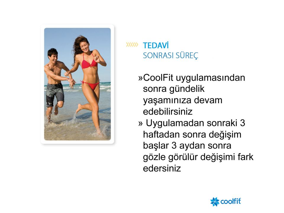
Tedavi sürerken sürekli hastanın yanında olunmasına gerek yoktur
Zeltiq vakum aplikatörü yan bölgeye uygulanmış

Benzer bir sunumlar


>")


