Sunuyu indir
Sunum yükleniyor. Lütfen bekleyiniz

1
Medical Instrumentation Application and Design, 4th Edition John Wiley and Sons Ltd, Feb 2009, Pages: 713 Basic Concepts of Medical Instrumentation Basic Sensors and Principles Amplifiers and Signal Processing The Origin of Biopotentials Biopotential Electrodes Biopotential Amplifiers Blood Pressure and Sound Measurement of Flow and Volume of Blood Measurements of the Respiratory System Chemical Biosensors Clinical Laboratory Instrumentation Medical Imaging Systems Therapeutic and Prosthetic Devices Electrical Safety KL-720_Intrduction0501 KL-720 Biomedical Measurement System (PPT) Laboratuvar Deneyleri
 Laboratuvar Deneyleri.")
2
KL-720 - Biyomedikal Ölçüm Sistemi

13
100mA Log scale

14
Ödevler: Bioelektrik empedans tanımı, ölçmeleri, dijital terazide vücudun yağ, su değerlerinin belirlenmesi Thermocouples Thermistor Resistance Temperature Detector Infrared Thermometers Pressure Transducers Load Cells Strain Gage Flowmeters pH Measurement Level Measurement 4 Kasım 2011 28 Ekim 2011

31
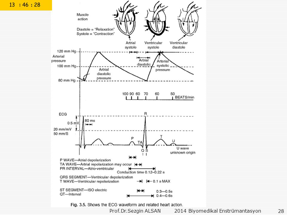
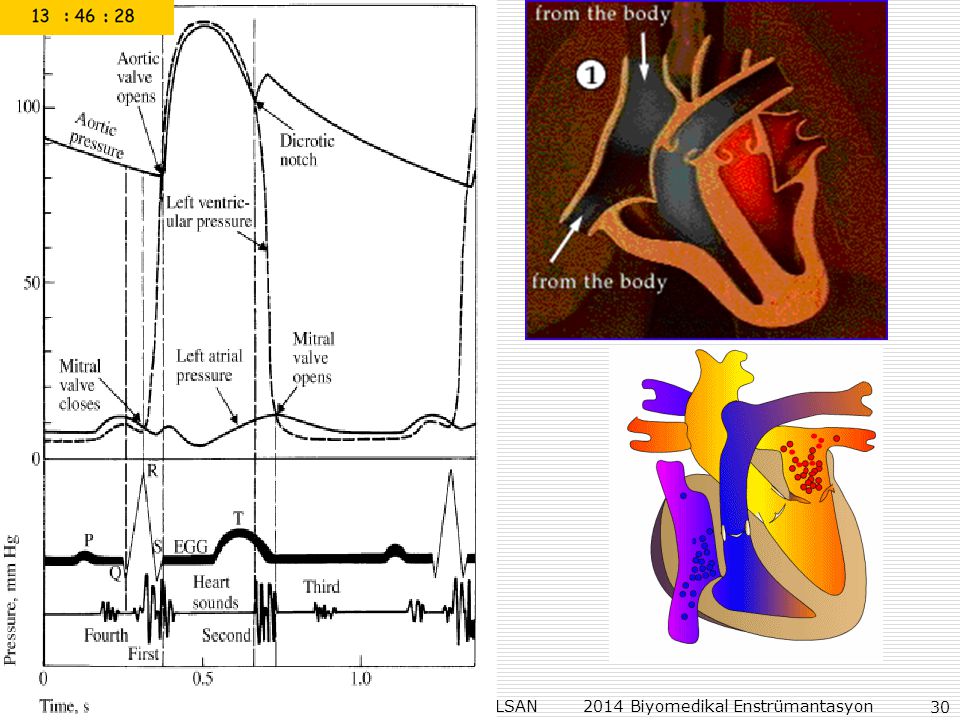
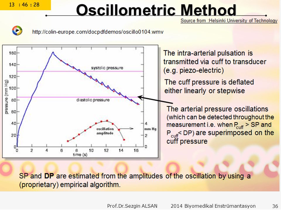
cardiac cycle Systole Diastole pressure volume flow

35
Elektrik akımı ve geriliminin insan için zararlı, öldürücü olma değerlerini, frekans özelliklerini de göz önüne alarak anlatın.(%20) Duyarlık elemanlarımızın cevaplarının zaman göre çıkışlarının: (giriş çıkış arasındaki gecikme var/yok, gecikmenin türü) zero-order (%5) firs-order (%5) second-order (%5) olma özelliklerini anlatın. EKG işaretlerinin algılanmasını, elde edilen işaretin genlik ve frekans özelliklerini, şeklini ve anlamlarını anlatın. (%20) Kan basıncını (mekanik-analog) ölçme yöntemini (ölçülen değerlerin anlamlarını belirterek) anlatın. (%15) Bioelektrik empedans tanımı, ölçmeleri, dijital terazide vücudun yağ, su değerlerinin belirlenmesi anlatın. (%20) Sadece üst tarafından erişilebilen 10 m derinlikte bir havuzunun içindeki suyun seviyesiini en güvenli şekilde ölçmek, ve display etmek için gerekli farklı iki sistemi tasarlayı, çizin, anlatın. Öneri-1(%5), Öneri-2(%5),
 zero-order (%5) firs-order (%5) second-order (%5) olma özelliklerini anlatın. EKG işaretlerinin algılanmasını, elde edilen işaretin genlik ve frekans özelliklerini, şeklini ve anlamlarını anlatın. (%20) Kan basıncını (mekanik-analog) ölçme yöntemini (ölçülen değerlerin anlamlarını belirterek) anlatın. (%15) Bioelektrik empedans tanımı, ölçmeleri, dijital terazide vücudun yağ, su değerlerinin belirlenmesi anlatın. (%20) Sadece üst tarafından erişilebilen 10 m derinlikte bir havuzunun içindeki suyun seviyesiini en güvenli şekilde ölçmek, ve display etmek için gerekli farklı iki sistemi tasarlayı, çizin, anlatın. Öneri-1(%5), Öneri-2(%5),")
38
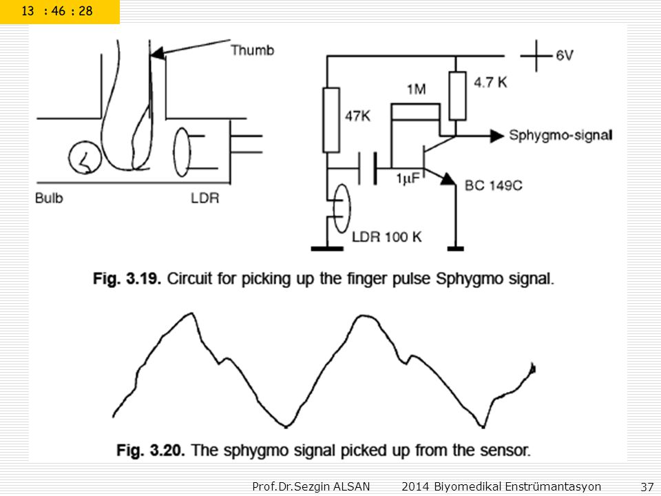
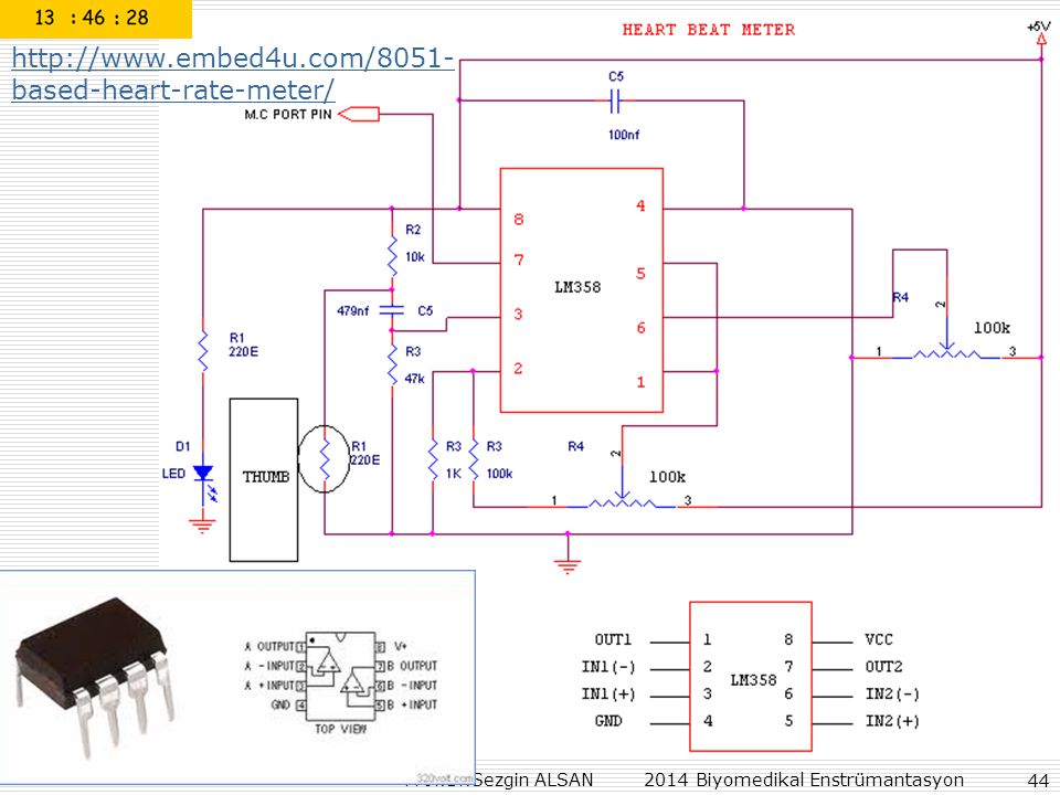
Heart rate: The sensor used in this board is TCRT1000, which is a reflective optical sensor with both the infrared light emitter and phototransistor placed side by side and are enclosed inside a leaded package so that there is minimum effect of surrounding visible light. The circuit diagram below shows the external biasing circuit for the TCRT1000 sensor. Pulling the Enable pin high will turn the IR emitter LED on and activate the sensor. A fingertip placed over the sensor will act as a reflector of the incident light. The amount of light reflected back from the fingertip is monitored by the phototransistor.

40
Miniature Wireless Pulse Oximeter
CMS-P PC Based Pulse Oximeter

42
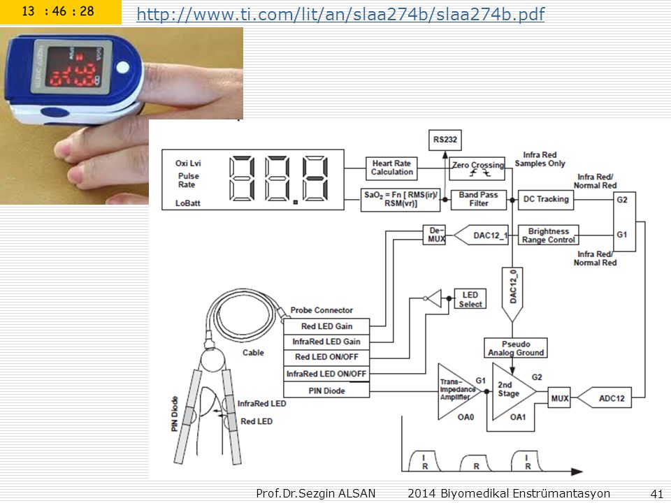
Pulse oximetry The probe contains two high intensity, monochromatic, light- emitting diodes, one emitting red light (660 nm) and the second infrared (940 nm) on one side and a photodetector on the other to measure the amount of light transmitted through the finger.
 and the second infrared (940 nm) on one side and a photodetector on the other to measure the amount of light transmitted through the finger.")
59
Ödevler: Bioelektrik empedans tanımı, ölçmeleri, dijital terazide vücudun yağ, su değerlerinin belirlenmesi Thermocouples Thermistor Resistance Temperature Detector Infrared Thermometers Pressure Transducers Load Cells Strain Gage Flowmeters pH Measurement Level Measurement

60
FREQUENCY MEASUREMENTS
ANALOG FREQUENCY MEASUREMENTS

61
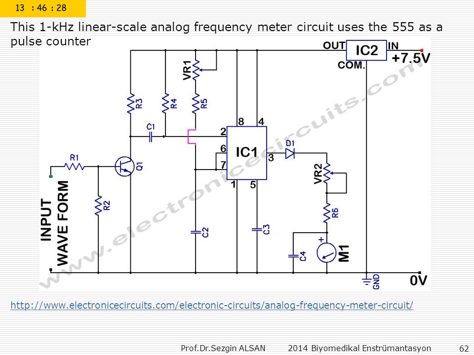
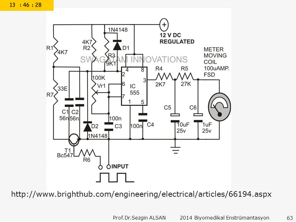
This audio frequency meter uses 555 IC as a monostable multivibrator (one-shoot trigger). A monostable multivibrator can act as a frequency-to-voltage converter because it produce a fixed pulse width, with the repetition rate/density is proportional to the triggering input frequency. Here is the circuit’s schematic diagram: For resistor R1, because it set the measurement range, it’s better to use a rotary switch to select different values for different ranges. For the ampere meter, you can use both analog or digital ampere meter. A cheap dual-slope ADC digital meter is suitable because its averaging characteristic, but a fast digital multimeter can also be used although it may show some uncertainty because of their fast sampling.

62
This 1-kHz linear-scale analog frequency meter circuit uses the 555 as a pulse counter
64
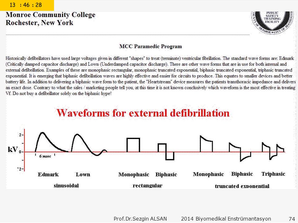
DEFIBRILLATORS For this example, the discharge is underdamped (biphasic, also referred to as a Gurvich waveform) when the patient resistance is less than about 56Ω because Rpatient+Rinductor=56Ω+10Ω=66Ω<Rcritical=67Ω In this case, the waveform is underdamped and produces a biphasic (oscillating) waveform. If the patient impedance is higher than 67Ω, the waveform is overdamped (monophasic, also referred to as an Edmark waveform). In this case the inductor slows the rate of rise of the discharge current, reduces the maximum voltage applied to the patient, and shapes the waveform to produce a damped sinusoidal waveform. The current delivered to the patient gradually rises to a rounded peak and drops back to zero. The discharge current pulse duration is about 2.5L1/C1, about 2.5 to 3.5ms for most defibrillators. DESIGN AND DEVELOPMENT OF MEDICAL ELECTRONIC INSTRUMENTATION
 when the patient resistance is less than about 56Ω because. Rpatient+Rinductor=56Ω+10Ω=66Ω<Rcritical=67Ω. In this case, the waveform is underdamped and produces a biphasic (oscillating) waveform. If the patient impedance is higher than 67Ω, the waveform is overdamped (monophasic, also referred to as an Edmark waveform). In this case the inductor slows the rate of rise of the discharge current, reduces the maximum voltage applied to the patient, and shapes. the waveform to produce a damped sinusoidal waveform. The current delivered to the. patient gradually rises to a rounded peak and drops back to zero. The discharge current. pulse duration is about 2.5L1/C1, about 2.5 to 3.5ms for most defibrillators. DESIGN AND DEVELOPMENT. OF MEDICAL ELECTRONIC. INSTRUMENTATION.")
65
Figure 8.32 Schematic diagram of a damped sinusoidal waveform defibrillator capable of delivering energies of up to 320 J into a 50-Ω patient load through a 5-ms Edmark (monophasic) waveform. Charge pushbutton SW2 energizes high-voltage transformer T1. C1 is charged through the high-voltage rectifier network D1–D4 and R1. Meter M1 is calibrated to yield an estimate of energy (in joules) delivered to the patient, assuming a load impedance of 50Ω. Defibrillation energy is delivered to the patient by simultaneously pressing on pushbuttons SW3 and SW4, which energize relay K1, which is used to transfer the defibrillation charge from capacitor C1 to the patient via pulse shaping inductor L1. R4 and R5 discharge C1 if the defibrillation buttons are depressed without a suitable load across the paddle electrodes or the
 waveform. Charge pushbutton SW2 energizes high-voltage transformer T1. C1 is charged through the high-voltage rectifier network D1–D4 and R1. Meter M1 is calibrated to yield an estimate of energy (in joules) delivered to the patient, assuming a load impedance of 50Ω. Defibrillation energy is delivered to the patient by simultaneously pressing on pushbuttons SW3 and SW4, which energize relay K1, which is used to transfer the defibrillation charge from capacitor C1 to the patient via pulse shaping inductor L1. R4 and R5 discharge C1 if the defibrillation buttons are depressed without a suitable load across the paddle electrodes or the.")
67
Energy limits A.C. defibrillator 0.25 sec × 230 volts × (I Amps) = 450 watts-sec. Required required I = 7.5Amps. But preferable 0.01 sec. Pulses. Then I = 450/(230 × 0.01) = 175 A. This is too much to be supplied by household mains.
 = 175 A. This is too much to be supplied by household mains.")
68
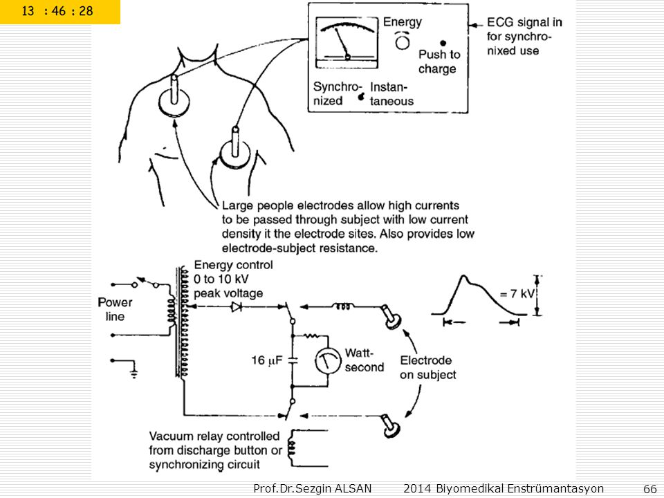
DC Defibrillation (Fig. 12.10b)
1. Safety housings for electrodes–capacitor discharges only when the electrodes are making firm contact with the heart or chest wall. 2. Two set of electrodes - not interchangeable sockets 1. Internal 50–72 J (5–3 kV) 3. Meter indicates Joules 1. External 400J (7 kV). 4. Charging time constant of 4 seconds M Ω × 16 μF 2. (charging resistor) Takes about 16 secs. to charge to 4 kV.
 3. Meter indicates Joules. 1. External 400J (7 kV). 4. Charging time constant of 4 seconds M Ω × 16 μF. 2. (charging resistor) Takes about 16 secs. to charge to 4 kV.")
69
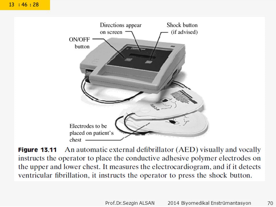
CAPACITIVE-DISCHARGE DC DEFIBRILLATORS
MEDICAL INSTRUMENTATION Application and Design FOURTH EDITION John G. Webster, Editor CAPACITIVE-DISCHARGE DC DEFIBRILLATORS A short high-amplitude defibrillation pulse can be obtained by using the capacitive-discharge circuit shown in Figure In this case, a half-wave rectifier driven by a step-up transformer is used to charge the capacitor C. A good rule of thumb is to keep charging time under 10 s.

71
When external electrodes are used, energies as high as 400 J may be
required. The energy stored in the capacitor is given by the well-known equation

72
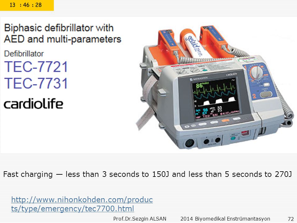
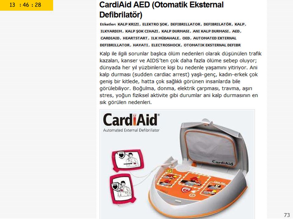
Fast charging — less than 3 seconds to 150J and less than 5 seconds to 270J
75
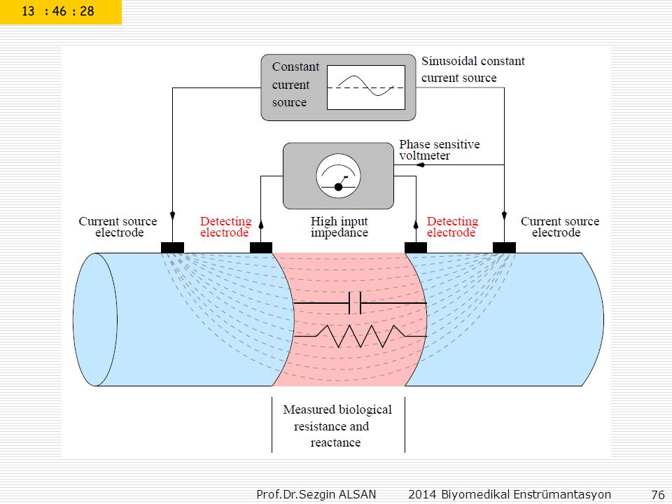
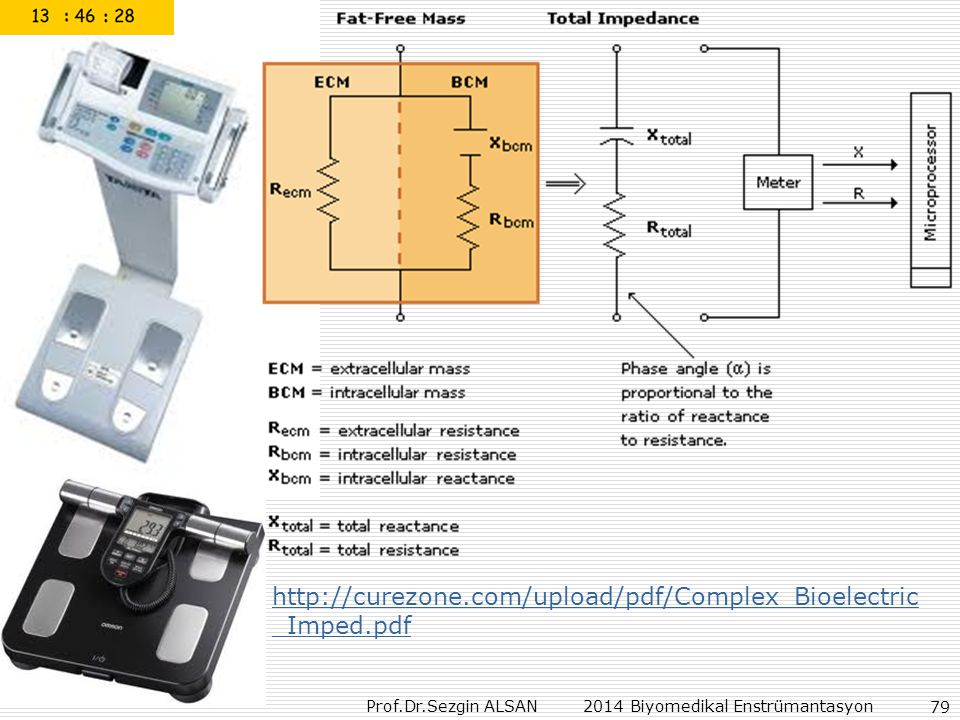
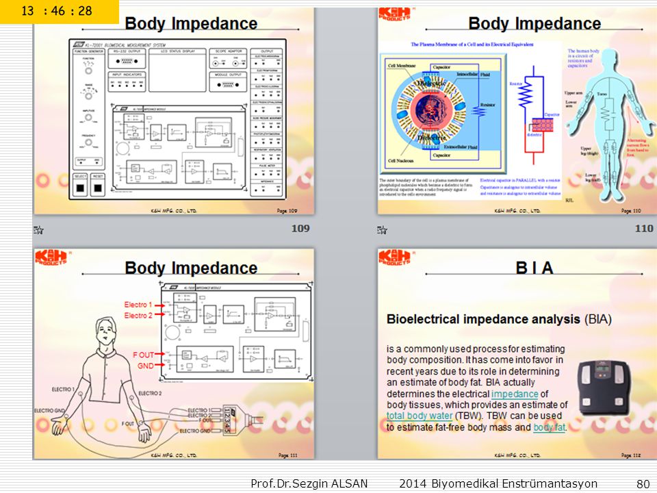
Principles of Bioelectrical Impedance Analysis

77
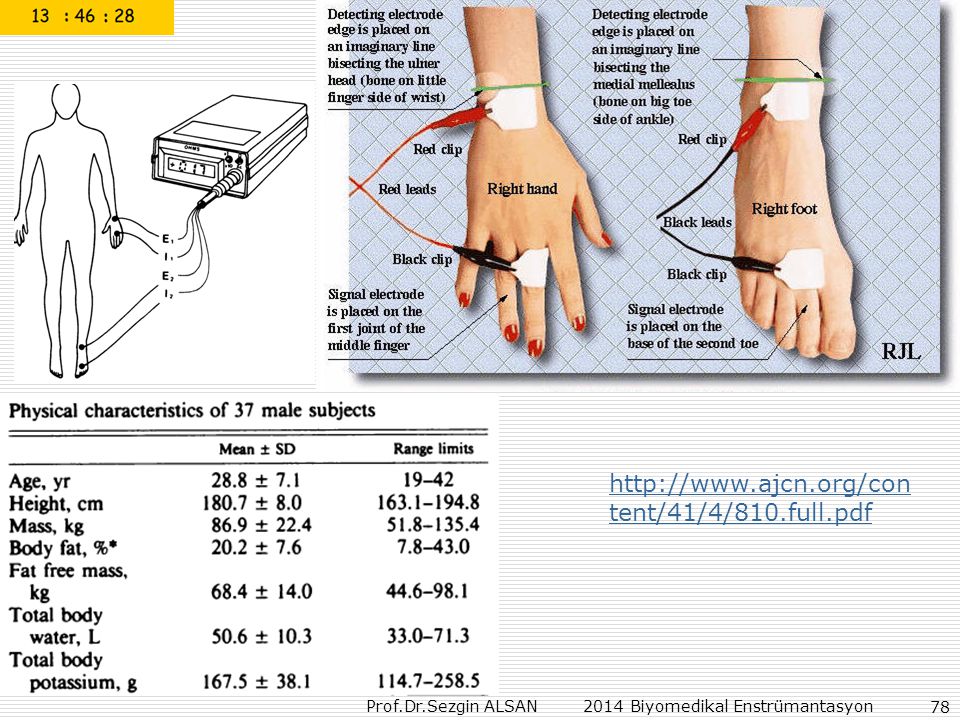
A method which involves the measurement of bioelectrical
resistive impedance (R) for the estimation of human body composition is described. This method is based upon the principle that the electrical conductivity of the fat-free tissue mass (FFM) is far greater than that of fat. Using an electrical impedance plethysmograph with a four electrode arrangement that introduces a painless signal (800 A at 50 kHz) into the body. FFM was assessed by hydrodensitometry and ranged from kg. Total body water (TBW) determined by D2O dilution and total body potassium (TBK) from whole body counting were 50.6 ± 10.3 L and 167.5 ± 38.1 g, respectively. .
 for the estimation. of human body composition is described. This method is based upon the principle that the electrical. conductivity of the fat-free tissue mass (FFM) is far greater than. that of fat. Using an electrical impedance plethysmograph with a four electrode. arrangement that introduces a painless signal (800 A at 50 kHz) into the body. FFM was assessed by hydrodensitometry and ranged. from kg. Total body water (TBW) determined by D2O dilution and total body. potassium (TBK) from whole body counting were 50.6 ± 10.3 L and ± 38.1 g, respectively. .")
81
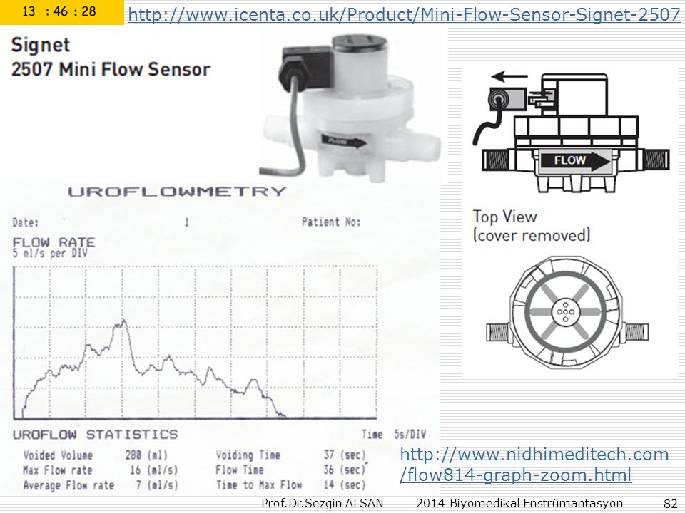
Uroflowmetry wireless
The SEDIA F1 Flowmeter provides you with a convenient, cost effective and easy to use solution for portable uroflowmetry. Communication between the flow meter and your computer is via Bluetooth connection. The SEDIA software is easily installed on your Windows PC, allowing you to print reports from your own printer. Tepe idrar akım hızı erkeklerde 20 ml/sn, kadınlarda ise 25 ml/sn üstünde normal oalrak kabul edilir

83
Turbine Flowmeters for Liquid Measurement

84
Peak-Flow Metre (Nefes ölçüm testi)
")
85
Peak-Flow Metre (Nefes ölçüm testi)
")
87
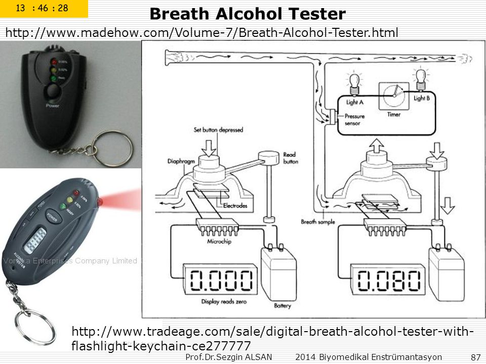
Breath Alcohol Tester
88
MQ303A is semiconductor sensor is for Alcohol detection,
Alcohol Tester Sensor MQ303A is semiconductor sensor is for Alcohol detection, Model No. MQ303A Sensor Type Semiconductor Standard Encapsulation Metal Detection Gas Alcohol Concentration ppm Alcohol Standard Circuit Conditions Heater Voltage V H 0.9V ± 0.1V AC or DC Loop Voltage Vc ≤6V DC Load Resistance R L Adjustable Heater Resistance R H 4.5W ± 0.5 W(Room Tem.) Heater Current IH 120±20mA Heater Power PH ≤ 140 mW Character Sensor Consumption PS ≤10 mW Sensing Resistance Rs 4KΩ-400KΩ(in air) Sensitivity S Rs(in air)/Rs(125ppm Alcohol)≥3 Slope α 0.50 ± 0.15(R300ppm/R100ppm Alcohol) Condition Tem. Humidity 20°C±2°C;65%±5%RH Standard test circuit Vc:3.0 V±0.1 V DC; VH: 0.9 V±0.1 V DC Preheat time Over 48 hours
 Heater Current. IH. 120±20mA. Heater Power. PH. ≤ 140 mW. Character. Sensor Consumption. PS. ≤10 mW. Sensing Resistance. Rs. 4KΩ-400KΩ(in air) Sensitivity. S. Rs(in air)/Rs(125ppm Alcohol)≥3. Slope. α ± 0.15(R300ppm/R100ppm Alcohol) Condition. Tem. Humidity. 20°C±2°C;65%±5%RH. Standard test circuit. Vc:3.0 V±0.1 V DC; VH: 0.9 V±0.1 V DC. Preheat time. Over 48 hours.")
89
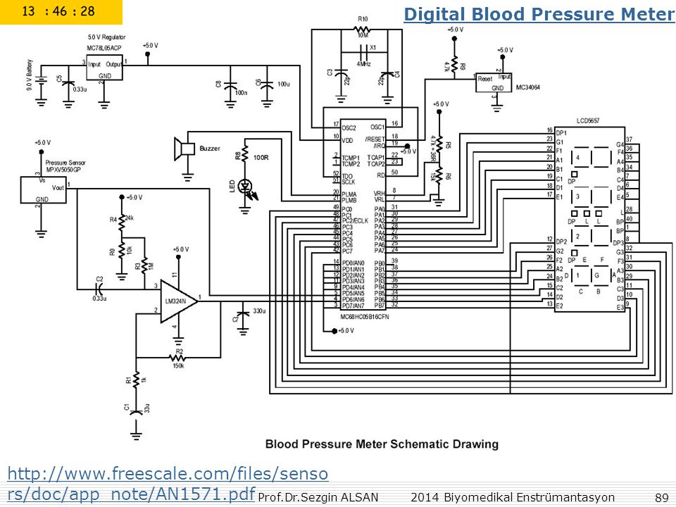
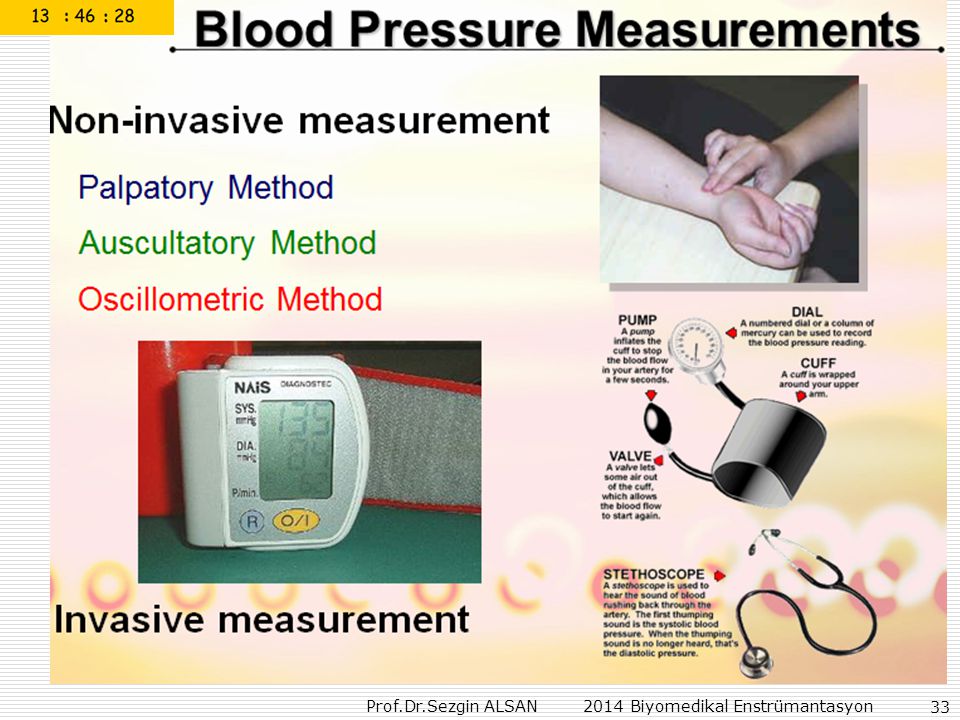
Digital Blood Pressure Meter
96
BİRİMİ ESKİ YENİ IŞINLANMA DOZU
TERİM BİRİMİ DÖNÜŞÜM ESKİ YENİ AKTİVİTE Curie (Ci) ; 3.7x10¹⁰ parçalanma / 1 saniye Becquerel (Bq); 1 parçalanma/1 saniye 1Ci=3.7x10¹⁰ Bq 1 Ci=37GBq IŞINLANMA DOZU Röntgen (R) ; normal hava şartlarında (00C ve 760 mm Hg basıncı) havanın 1kg’ında 2.58x10⁻⁴ Coulomb’luk elektrik yükü değerinde (+) ve (-) iyonlar oluşturan X veya g radyasyonu miktarıdır. Coulomb / kilogram (C/kg) ; normal hava şartlarında havanın 1 kg’ında 1 Coulomb’luk elektrik yükü değerinde (+) ve (-) iyonlar oluşturan X veya g radyasyonu miktarıdır. 1C/kg=3876 R1R=2.58x10⁻⁴ C/kg SOĞURULMUŞ DOZ radiation oz (rad); ışınlanan maddenin 1 kg’ında 10-2 Joule’lük enerji soğurulması meydana getiren herhangi bir radyasyon miktarıdır. Gray (Gy) ; ışınlanan maddenin 1 kg’ında 1 Joule’lük enerji soğurulması meydana getiren herhangi bir radyasyon miktarıdır. 1Gy=100rad 1rad=0.01 Gy DOZ EŞDEĞERİ röntgen equivalent man (); 1 Röntgenlik X veya ışını ile aynı biyolojik etkiyi oluşturan herhangi bir radyasyon miktarıdır. rem=(rad)x(WR)* Sievert (Sv) ; 1 Gy’lik X ve g ışını ile aynı biyolojik etkiyi meydana getiren herhangi bir radyasyon miktarıdır. Sv= (Gy)x(WR)* 1Sv=100 rem 1rem=0.01Sv
 ; 3.7x10¹⁰ parçalanma / 1 saniye. Becquerel (Bq); 1 parçalanma/1 saniye. 1Ci=3.7x10¹⁰ Bq 1 Ci=37GBq. IŞINLANMA DOZU. Röntgen (R) ; normal hava şartlarında (00C ve 760 mm Hg basıncı) havanın 1kg’ında 2.58x10⁻⁴ Coulomb’luk elektrik yükü değerinde (+) ve (-) iyonlar oluşturan X veya g radyasyonu miktarıdır. Coulomb / kilogram (C/kg) ; normal hava şartlarında havanın 1 kg’ında 1 Coulomb’luk elektrik yükü değerinde (+) ve (-) iyonlar oluşturan X veya g radyasyonu miktarıdır. 1C/kg=3876 R1R=2.58x10⁻⁴ C/kg. SOĞURULMUŞ DOZ. radiation oz (rad); ışınlanan maddenin 1 kg’ında 10-2 Joule’lük enerji soğurulması meydana getiren herhangi bir radyasyon miktarıdır. Gray (Gy) ; ışınlanan maddenin 1 kg’ında 1 Joule’lük enerji soğurulması meydana getiren herhangi bir radyasyon miktarıdır. 1Gy=100rad 1rad=0.01 Gy. DOZ EŞDEĞERİ. röntgen equivalent man (); 1 Röntgenlik X veya ışını ile aynı biyolojik etkiyi oluşturan herhangi bir radyasyon miktarıdır. rem=(rad)x(WR)* Sievert (Sv) ; 1 Gy’lik X ve g ışını ile aynı biyolojik etkiyi meydana getiren herhangi bir radyasyon miktarıdır. Sv= (Gy)x(WR)* 1Sv=100 rem 1rem=0.01Sv.")
97
Normal background radiation varies from place to place but delivers a dose equivalent in the vicinity of 2.4 mSv/year, or about 0.3 µSv/h. The international limit for radiation exposure for nuclear workers is 20 mSv per year, averaged over five years, with a limit of 50 mSv in any one year, however for workers performing emergency services EPA guidance on dose limits is 100 mSv when "protecting valuable property" and 250 mSv when the activity is "life saving or protection of large populations. A 250 mSv dose is estimated to increase one's lifetime risk of developing fatal cancer from about 20% to about 21%, and chronic exposure of 100 mSv per year is the "lowest level at which any increase in cancer is clearly evident," according to the International Commission on Radiological Protection. Symptoms of radiation poisoning typically emerge with a 1000 mSv total dose over a day.

98
Bazı radyolojik tetkikler sonucu, ülke seviyelerine ve yapılan tetkiklere göre, hastaların maruz kaldığı etkin dozlar. TETKİKLER HER BİR TETKİKTE MARUZ KALINAN ETKİN DOZ (mSv) Seviye 1* Seviye 2** Seviye 3-4*** Dünya Göğüs Radyografisi 0.14 0.20 Göğüs Fotofloroskopisi 0.65 Göğüs Floroskopisi 1.1 Kol,bacak ve eklemler 0.06 0.1 Omurga Bel 1.8 2 Göğüs 1.4 1.5 Boyun 0.27 0.3 Kalça ve Kalça eklemi 0.83 1 Kafa 0.15 Karın 0.5 0.6 0.55 Üst sindirim sistemi 3.6 4 3.7 Alt sindirim sistemi 6.4 Safra kesesi grafisi Üriner sistem grafisi 3.9 Mamografi Bilgisayarlı Tomografi 8.8 5 8.6 Anjiyografi 12 Cerrahi işlemler 20 Diş 0.02 0.03
 Seviye 1* Seviye 2** Seviye 3-4*** Dünya. Göğüs Radyografisi Göğüs Fotofloroskopisi Göğüs Floroskopisi Kol,bacak ve eklemler Omurga. Bel Göğüs Boyun Kalça ve Kalça eklemi Kafa Karın Üst sindirim sistemi Alt sindirim sistemi Safra kesesi grafisi. Üriner sistem grafisi Mamografi. Bilgisayarlı Tomografi Anjiyografi. 12. Cerrahi işlemler. 20. Diş")
100
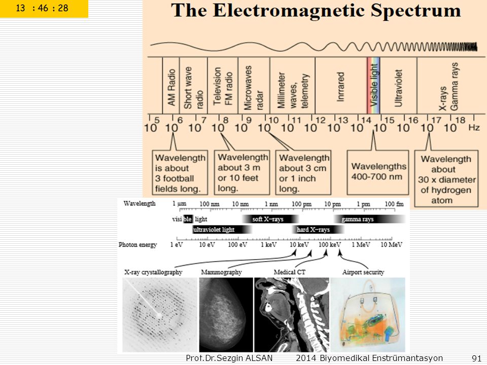
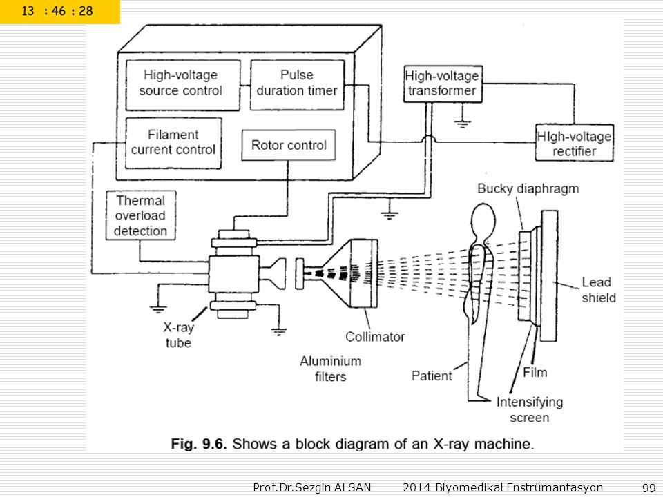
Computed tomography (CT) scanning, also called computerized axial tomography (CAT) scanning, is a medical imaging procedure that uses x-rays to show cross-sectional images of the body. A CT imaging system produces cross-sectional images or "slices" of areas of the body, like the slices in a loaf of bread. These cross-sectional images are used for a variety of diagnostic and therapeutic purposes. How a CT system works: 1.A motorized table moves the patient through a circular opening in the CT imaging system. A motorized table moves the patient through a circular opening in the CT imaging system. While the patient is inside the opening of the CT imaging system, an x-ray source and detector within the housing rotate around the patient. A single rotation takes about 1 second. The x-ray source produces a narrow, fan-shaped beam of x-rays that passes through a section of the patient's body. A detector opposite from the x-ray source records the x-rays passing through the patient's body as a "snapshot" image. Many different "snapshots" (at many angles through the patient) are collected during one complete rotation. For each rotation of the x-ray source and detector, the image data are sent to a computer to reconstruct all of the individual "snapshots" into one or multiple cross-sectional images (slices) of the internal organs and tissues.
 are collected during one complete rotation. For each rotation of the x-ray source and detector, the image data are sent to a computer to reconstruct all of the individual snapshots into one or multiple cross-sectional images (slices) of the internal organs and tissues.")
Benzer bir sunumlar


 27.03.2008.>")


 “HİZMET MEMNUNİYETİ ÇALIŞMASI” Temmuz, 2010.>")
